
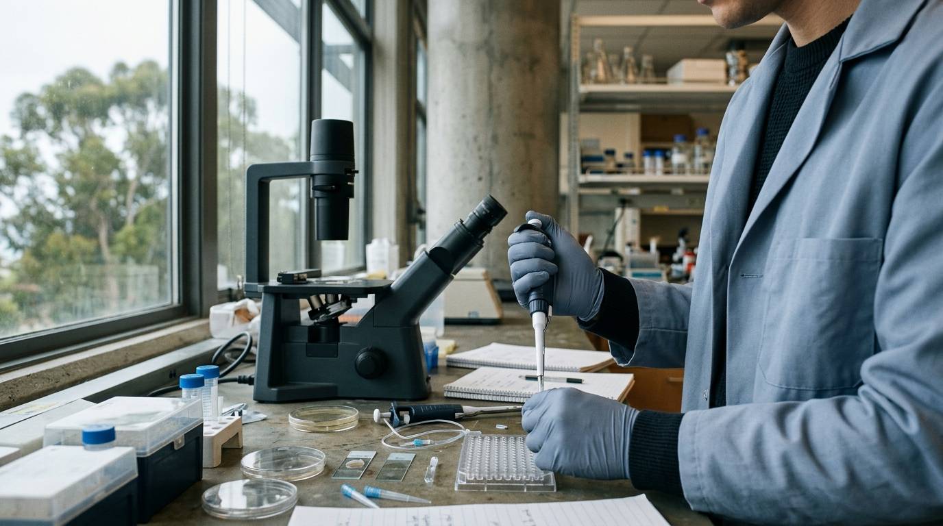
More blood vessels do not always equal better tissue recovery. In experimental biology, researchers often assume that if a compound triggers vessel sprouting, the tissue has healed. Yet structural vessel presence and active tissue perfusion are distinct physiological states. A dense network of capillaries visible under a microscope can remain nonfunctional if blood fails to circulate through the new lumens.
Understanding tissue repair requires looking past static histology. Researchers must measure how new microvascular networks assemble, whether endothelial cells communicate correctly, and if newly sprouted vessels actually carry circulation.
Define the Difference Between Vascular Growth and Tissue Recovery
Angiogenesis describes the biological process through which new blood vessels sprout from preexisting vasculature. In tissue recovery research, investigators study how vascular remodeling restores oxygen and nutrients to damaged cells. However, finding isolated endothelial cell markers does not guarantee tissue survival or restoration.
True repair requires functional microvascular networks that support hemodynamic flow. When assessing experimental compounds, evaluating preclinical research findings requires separating structural markers from functional recovery. An increase in capillary density without adequate lumen formation or pericyte coverage leaves tissue vulnerable to hypoxia. Investigating vascular endpoints therefore demands direct observation of vessel architecture alongside active blood flow over time.
Categorize Preclinical Evidence Levels in Microvascular Research
Evaluating angiogenesis requires understanding the tiers of experimental proof. Laboratory cell cultures show whether an agent promotes endothelial migration or proliferation. However, in vitro assays cannot replicate systemic pressure, shear stress, or matrix resistance.
- Preclinical Evidence Hierarchy
- In vitro cell migration and tube formation assays
- Ex vivo tissue explant outgrowths
- In vivo animal models with static histology
- In vivo longitudinal imaging with functional perfusion tracing
Animal research provides systemic context, but static post-mortem histology captures only a single, frozen moment. As described in frameworks for classifying research evidence, animal findings cannot be directly assumed to reflect human biology. The most rigorous preclinical platforms use longitudinal in vivo tracking in living organisms. These live imaging setups allow researchers to observe vessel formation across multiple stages of repair.
Select Appropriate Imaging Models for In Vivo Angiogenesis
Evaluating microvascular remodeling requires models that preserve native tissue architecture. Traditional wound repair assays often rely on histological sectioning, which damages delicate capillary sprouts and eliminates dynamic information.
To overcome this limitation, researchers developed a minimally invasive wound healing model in the mouse ear. Published in Scientific Reports, this animal model utilized mice expressing GCaMP2 in arterial endothelial cells. The thin tissue of the mouse ear allowed researchers to deploy two-photon microscopy with high spatiotemporal resolution.
This model tracked arterial sprouts over time in the same living animal without repeated surgical intervention. By maintaining an intact physiological setting, researchers could record cell behavior as the wound microenvironment evolved.
Trace Endothelial Calcium Signals and Growth Factor Dynamics
Sprouting angiogenesis begins when specialized endothelial cells detect biochemical gradients released by injured tissue. In the mouse-ear wound model, researchers observed that arterial endothelial sprouts developed reliably after injury.
The investigators recorded calcium activity within arterial endothelial cells during this regenerative response. These intracellular calcium transients were more numerous on the distal segment of the injured tissue. The observed calcium activity was consistent with vascular endothelial growth factor stimulation.
Tracking these ionic signals demonstrates the dynamic nature of endothelial activation. Similar to differentiating initial signaling from downstream physiological outcomes, intracellular calcium surges indicate receptor engagement. However, these signaling events mark only the beginning of a complex cellular sequence.
Quantify Functional Perfusion Versus Structural Vessel Presence
The definitive test of a blood vessel is its ability to conduct blood. In the Cx40-GCaMP2 mouse model, researchers tested vessel functionality by combining two-photon microscopy with an intravenously administered fluorescent tracer.
The team administered the intravascular tracer Texas Red to visualize active plasma flow through newly formed vessels. By day eight after injury, proximal and distal arteries were structurally connected across the wound bed. Yet functional assessment revealed a striking mismatch between structure and circulation.
Although the sprouts formed physical bridges across the injured zone, only two-thirds of the detected sprouts were actually perfused at day eight. The remaining one-third of the vessels were physically visible but lacked functional circulation. This audited finding underscores why surrogate structural metrics can mislead investigators. Counting sprouts alone overestimates the true circulatory capacity of the healing tissue.
Identify Methodological Limitations and Translational Gaps
Every experimental system has clear biological boundaries. The mouse-ear two-photon model provides high spatial resolution, but it uses specialized transgenic rodents under controlled laboratory conditions. The thin, cartilage-rich environment of an ear differs markedly from deep visceral organs or human dermal tissue.
Furthermore, data from mouse-ear models represent preclinical animal evidence. These findings cannot establish efficacy, dosing, or therapeutic outcomes in human clinical medicine. Transgenic markers such as GCaMP2 and Cx40 reporters also introduce specific observation biases toward arterial subsets.
Researchers must also account for timeline limitations. Observing vessels at day eight captures an intermediate remodeling phase rather than final tissue stabilization. Unperfused sprouts might mature and carry blood later, or they could regress through natural vessel pruning.
Apply Analytical Standards to Vascular Research Reagents
Investigating microvascular signaling pathways requires well-characterized laboratory reagents. Peptides and small molecules used to study angiogenic mechanisms must meet rigorous chemical verification standards before use in experimental protocols.
When reviewing laboratory documentation, verifying experimental reagent quality is essential for experimental reproducibility. A Certificate of Analysis demonstrates only what analytical testing detected in a specific sample. It does not certify stability across varying laboratory environments or predict biological performance in vivo.
Laboratories rely on High-Performance Liquid Chromatography and Mass Spectrometry to confirm chemical identity and purity. As highlighted in rigorous analytical standards for peptide purity, relying on a single analytical trace can overlook residual salts or synthesis impurities. Verifying raw materials ensures that measured endothelial responses stem from the target compound rather than synthesis artifacts.
Key Takeaways
- Angiogenesis involves both structural sprout formation and the development of functional blood circulation.
- Two-photon microscopy in mouse-ear wound models allows longitudinal tracking of individual arterial sprouts in living tissue.
- Endothelial calcium transients observed after tissue injury align with growth factor stimulation patterns during vessel outgrowth.
- Structural vessel connection does not guarantee blood flow; experimental tracking showed only two-thirds of connected sprouts were perfused at day eight.
- Preclinical imaging metrics from animal models cannot be extrapolated to prove human therapeutic outcomes.
Accurate tissue repair research demands that scientists measure functional perfusion rather than relying solely on visual vessel counts.
Sources
Introducing / CaliLean Field Notes
A little more
perspective.
An editorial letter for curious minds. Thoughtful reading, questions worth keeping and the sources behind them.
Explore Field Notes Join the list for the first edition

