
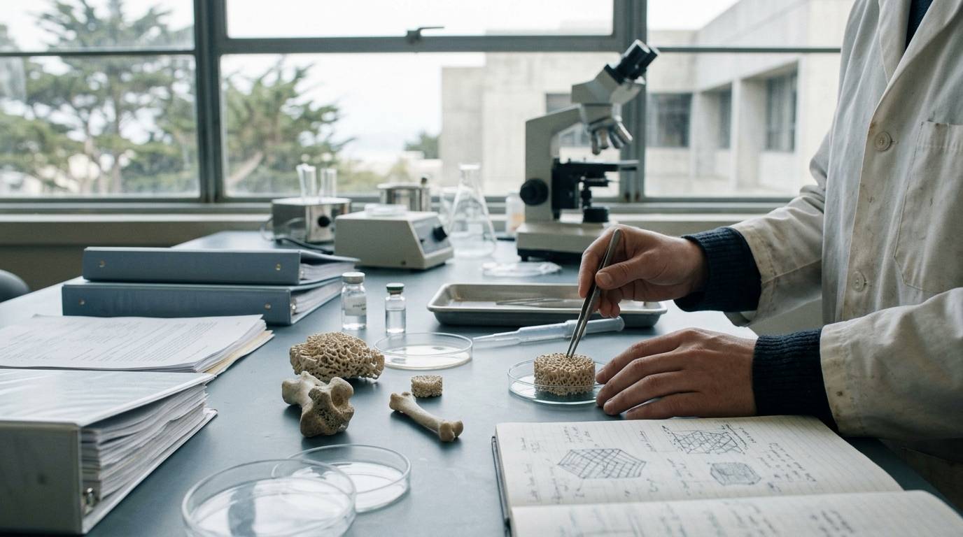
Bone repair is often assumed to be a continuous process of mineral accumulation. In reality, successful regeneration relies equally on active tissue breakdown to reshape provisional bone calluses. Assessing this process requires researchers to separate simple mineral deposition from true load-bearing recovery. This guide examines the cellular remodeling mechanisms, vascular requirements, and analytical testing methods that define bone repair research.
Preclinical bone research relies heavily on structural imaging, tissue staining, and mechanical validation. Interpreting these study designs requires understanding the distinct roles of osteoblasts, osteoclasts, and blood supply. It also requires understanding the predictive value and limits of micro-computed tomography.
Distinguish Cellular Turnover from Structural Deposition
Bone healing is not a static calcification event. It is a dynamic exchange between matrix formation and resorption. Research into these cellular dynamics requires evaluating both osteoblast-mediated bone synthesis and osteoclast-mediated remodeling. When looking at biological pathways and cellular mechanisms, the timing of cellular recruitment determines whether a fracture site successfully bridges.
Specific receptor pathways directly alter this cellular timeline. In an original animal study published in the Journal of Bone and Mineral Research, investigators evaluated closed stabilized femoral fractures in mice lacking the EP1 prostanoid receptor alongside wild-type controls. The researchers tracked histological markers across the healing period.
At day 14 post-fracture, the EP1 knockout mice displayed a higher presence of tartrate-resistant acid phosphatase-positive osteoclasts in the callus. This early increase in osteoclast activity accompanied accelerated bone remodeling. The knockout model demonstrated an earlier return to normal bone morphometry compared to wild-type controls.
These findings illustrate that faster bone healing does not depend solely on accelerated mineral addition. Instead, the timely activation of resorption pathways allows the provisional callus to resolve into organized cortical structure. Preclinical models must therefore monitor both bone-forming cells and catabolic remodeling markers.
Trace Vascular Pathways into the Regenerating Callus
Mineralization cannot occur in an isolated environment. The regenerating fracture site is metabolically demanding and requires continuous perfusion. According to a scientific review published in Current Osteoporosis Reports, revascularization serves multiple physiological functions during bone repair.
The review notes that new blood vessels supply oxygen and essential nutrients to the highly active regenerating callus. Without adequate oxygen delivery, local progenitor cells cannot sustain the metabolic output needed for tissue synthesis. Neovascularization prevents tissue necrosis within the hypoxic core of the injury.
Beyond nutrient delivery, newly formed vessels serve as physical conduits for cellular traffic. The literature indicates that blood vessels provide a pathway for inflammatory cells to reach the damaged area. These vessels also allow cartilage-precursor and bone-precursor cells to migrate into the repair zone.
While literature reviews summarize these established biological principles, they synthesize broad observations rather than measuring a single experimental variable. Researchers establishing causality must evaluate these transport pathways in primary experimental settings. Preclinical studies should confirm whether structural outcomes stem from direct osteogenic signaling or secondary vascular support.
Correlate Micro-CT Structural Data with Functional Biomechanics
Quantifying bone regeneration requires methods that capture both structural geometry and functional competence. Micro-computed tomography (micro-CT) provides three-dimensional, high-resolution assessments of mineralized tissue. However, researchers must verify whether radiographic density reflects true mechanical strength.
An original animal study published in the Journal of Orthopaedic Research evaluated this relationship directly. Investigators examined 188 murine fracture calluses across multiple post-fracture time points. The study applied different experimental conditions designed to alter fracture healing rates, combining structural imaging with destructive mechanical testing.
The researchers paired micro-CT evaluations directly with torsional mechanical testing on the same specimens. Their abstract reports that several micro-CT-derived measures of callus structure and composition predicted the mechanical properties of the callus. Structural dimensions and mineral distribution correlated with the load-bearing capacity of the repairing bone.
These data demonstrate that high-resolution imaging can serve as a dependable surrogate for structural stability in rodent models. When researchers evaluate interpreting preclinical peptide studies, pairing imaging with physical load tests provides stronger evidence than imaging alone. Radiographic mineralization must translate into mechanical competence to indicate true tissue recovery.
Account for Model Limitations and Measurement Gaps
Preclinical bone repair research carries methodological limitations that restrict immediate human translation. Rodent fracture models provide controlled genetic environments, such as the EP1 knockout framework. However, rodent bone biology differs from human skeletal physiology in critical ways.
Small rodents possess different cortical structures and lack the Haversian remodeling systems found in larger mammals. Stabilized fractures in mice also experience different biomechanical forces than human skeletal injuries. Torsion testing provides reliable data on shear resistance, but it does not reproduce the complex multiaxial loads experienced during clinical weight-bearing.
Review articles describe the general necessity of vascular networks, but they do not prove that augmenting blood flow alone accelerates healing in every context. Preclinical studies must clearly identify their evidence tier. Frameworks outlined in evidence hierarchy frameworks emphasize that animal data and narrative reviews cannot serve as proof of clinical efficacy.
Furthermore, micro-CT imaging detects mineral density, but it cannot measure cellular viability or tissue compliance on its own. A callus may appear dense and mineralized while harboring non-viable fibrous tissue or residual cartilage remnants. Destructive mechanical testing confirms structural strength, but it only reflects a single time point in a sacrificed animal model.
Evaluate Laboratory Controls and Analytical Boundaries
Rigorous regenerative research depends on verified experimental materials and documented testing standards. Preclinical investigators studying bioactive molecules or matrix scaffolds must confirm the exact identity and purity of their research compounds. Analytical validation protects experimental models from confounding artifacts caused by degradation or residual synthesis impurities.
Standard laboratory documentation, such as a Certificate of Analysis (COA), verifies the biochemical metrics of a specific material sample. As detailed in resources covering analytical laboratory testing, a COA confirms the chemical characterization of a submitted batch through methods like mass spectrometry and liquid chromatography.
However, analytical verification carries specific boundaries:
- A COA confirms physical identity and purity for the tested sample only.
- It does not guarantee the stability of the compound across varying storage conditions.
- Analytical purity metrics do not predict biological uptake, tissue clearance, or receptor binding in an animal model.
- Chemical documentation cannot prove therapeutic safety or clinical efficacy.
Maintaining precise laboratory boundaries ensures that preclinical findings remain grounded in reproducible science. Documented testing standards provide the material control necessary to evaluate bone remodeling pathways without interference from variable material quality.
Key Takeaways
- Bone regeneration requires both osteoblast-mediated matrix deposition and osteoclast-mediated tissue resorption.
- EP1 knockout mice exhibit increased osteoclast presence at day 14, leading to accelerated callus remodeling.
- Vascular ingrowth brings oxygen, nutrients, inflammatory cells, and skeletal precursor cells directly into the fracture site.
- Micro-CT metrics of callus structure and composition can reliably predict torsional mechanical properties in murine models.
- Preclinical rodent experiments provide structural and mechanistic insight, but they cannot replace controlled human clinical trials.
- Analytical testing documents compound purity for specific batches, but it does not establish biological safety or clinical efficacy.
Precise bone regeneration research relies on combining cellular markers, vascular tracking, and mechanical validation rather than relying on structural imaging alone.
Sources
Introducing / CaliLean Field Notes
A little more
perspective.
An editorial letter for curious minds. Thoughtful reading, questions worth keeping and the sources behind them.
Explore Field Notes Join the list for the first edition

