
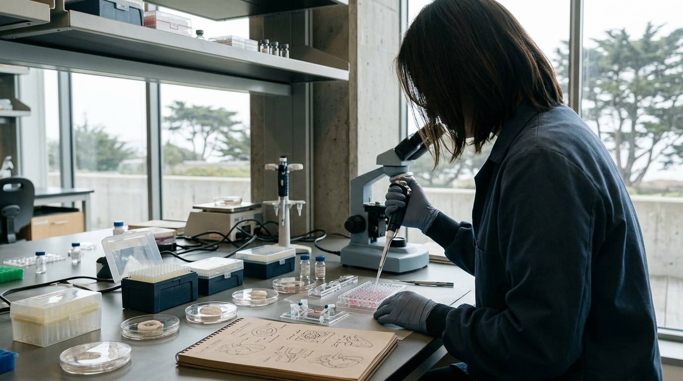
A laboratory team examines tissue sections following an experimental cardiac injury protocol. The assay shows alterations in extracellular collagen and marked shifts in local cell density. It is tempting to describe these structural shifts as myocardial regeneration. In published cardiac biology, researchers must carefully separate replacement scar formation from actual parenchymal regeneration.
This guide outlines how cardiac research separates functional scar formation from genuine tissue regeneration. It reviews the biological definitions of repair, the specific experimental animal models used in published literature, and the strict evidence standards required to interpret post-injury cardiac data.
Defining cardiac repair versus cardiac regeneration
Cardiac injury outcomes exist along a clear spectrum between reparative scar stabilization and genuine myocardial renewal. In adult mammalian biology, the primary response to ischemic necrosis is wound healing via fibrosis. This response maintains ventricular wall integrity, but it does not replace lost contractile tissue.
True regeneration requires the proliferation of functional cardiomyocytes and the restoration of normal myocardial architecture. In contrast, repair involves the expansion of non-contractile cells, extracellular matrix deposition, and scar maturation. Conflating these two distinct biological processes is a frequent issue in preclinical literature.
When evaluating experimental findings, researchers must distinguish between in vitro assays, controlled animal interventions, and observational human tissue samples. Research in regenerative research evaluations demonstrates that cellular differentiation markers observed in cell cultures cannot confirm organ-level functional recovery. Controlled animal studies remain necessary to track cell lineage and tissue-level architecture over time.
Observational examinations of human cardiac tissue can confirm that specific cellular states exist in clinical scars. However, static human tissue samples cannot establish cell origins or prove dynamic differentiation trajectories. True mechanistic verification requires rigorous, lineage-traced animal models.
How experimental animal models differ
The choice of experimental model dictates whether cardiac tissue responds with regenerative proliferation or fibrotic scar repair. A concrete example of why model selection matters comes from original research comparing neonatal mice at different developmental stages.
In a published surgical model, one-day-old neonatal mice underwent surgical removal of part of the ventricular apex. The hearts of these one-day-old mice demonstrated cardiomyocyte proliferation with minimal hypertrophy or fibrosis. This response allowed true regeneration of the resected tissue.
When investigators tested the exact same apical resection protocol in seven-day-old mice, this regenerative capacity was completely absent. The older neonatal hearts responded with fibrotic repair rather than parenchymal proliferation. This sharp biological divergence highlights the risk of assuming all animal models reflect adult human biology.
Adult animal models rely predominantly on myocardial infarction protocols, including coronary artery ligation. In adult mice, resident non-cardiomyocyte populations govern the injury response. Applying insights from transient neonatal regenerative windows to mature adult cardiac repair misrepresents the underlying biology. Careful researchers review the precise developmental stage before evaluating preclinical study designs.
Cellular mechanisms of post-injury scar formation
Scar formation in adult cardiac tissue follows a defined cellular and temporal sequence driven by resident cardiac fibroblasts. Genetic lineage-tracing experiments show that non-cardiomyocyte populations govern each phase of healing. The tissue response progresses through distinct stages of activation, structural support, and long-term stabilization.
Following an acute myocardial infarction, resident cardiac fibroblasts become activated and proliferate rapidly. These fibroblasts reach their maximum rate of proliferation within two to four days after the infarction injury. During this initial window, the resident fibroblast population expands 3.5-fold and remains sustained in the tissue long term.
Between three and seven days after injury, activated fibroblasts differentiate into mature myofibroblasts. These myofibroblasts express smooth muscle alpha-actin and secrete abundant extracellular matrix proteins. This matrix deposition provides necessary structural support to reinforce the necrotic ventricular wall.
Between 7 and 10 days post-infarction, the cellular dynamics shift again. The myofibroblasts lose their proliferative capacity and cease smooth muscle alpha-actin expression. The surrounding collagen matrix matures into a stable scar, and the persisting lineage-traced cells enter a specialized differentiated state termed a matrifibrocyte.
Key findings from lineage tracing and resection studies
Rigorous primary studies establish clear empirical boundaries for what adult mammalian cardiac tissue can accomplish after injury. Rather than demonstrating de novo cardiomyocyte creation, lineage-traced adult models show a tightly controlled fibrotic sequence.
The published evidence documents several verified cellular findings:
- Resident cardiac fibroblasts expand 3.5-fold within two to four days following myocardial infarction injury in mice.
- Proliferating fibroblasts differentiate into smooth muscle alpha-actin-positive myofibroblasts between three and seven days post-injury to support the necrotic zone.
- Myofibroblasts lose smooth muscle alpha-actin expression and stop proliferating between 7 and 10 days as the scar fully matures.
- Initial lineage-traced fibroblasts persist indefinitely in the mature scar as specialized matrifibrocytes.
- Cardiomyocyte proliferation and minimal fibrosis occur after partial apical resection in one-day-old mice, but this capacity is completely lost by seven days of age.
These findings show that adult myocardial tissue focuses on preserving wall geometry through specialized extracellular matrix production. Studies tracking molecular pathways must separate this structural adaptation from contractile renewal. Readers can review binding models versus functional biological outcomes to see how cellular assays differ from physiological recovery.
Common mistakes when interpreting cardiac repair data
The most pervasive mistake in interpreting cardiac research is treating reduced scar size or matrix shifts as proof of muscular regeneration. Interventions that modify fibroblast behavior can alter scar geometry without generating any new contractile tissue. Evidence of tissue remodeling should never be assumed to indicate new cardiomyocyte formation.
Another critical error is conflating observational human tissue data with interventional proof. Investigators have identified cells resembling matrifibrocytes within scarred human myocardium. However, observing these cells in clinical specimens does not turn the original study into a controlled human clinical trial. The lineage-tracing proof demonstrating that these cells arise from resident cardiac fibroblasts was performed entirely in mice.
Translating findings across developmental windows is equally problematic. Demonstrating cardiomyocyte proliferation in a one-day-old neonatal mouse does not indicate that mature adult mammalian hearts possess the same regenerative capacity. The complete disappearance of that regenerative ability by day seven underscores the extreme age-dependence of cardiac plasticity.
Finally, researchers must evaluate research materials based on verified laboratory testing standards. A Certificate of Analysis confirms only the chemical purity and identity of a specific synthesized compound batch. It does not provide evidence of tissue survival, in vivo efficacy, or biological activity in cardiac models.
When to revisit this resource
Revisit this resource when designing cardiac injury models, evaluating published literature that reports cardiac regeneration, or auditing endpoints in tissue repair studies. A clear distinction between scar maturation and true tissue regeneration ensures that research conclusions remain grounded in verified experimental evidence.
Sources
Introducing / CaliLean Field Notes
A little more
perspective.
An editorial letter for curious minds. Thoughtful reading, questions worth keeping and the sources behind them.
Explore Field Notes Join the list for the first edition

