
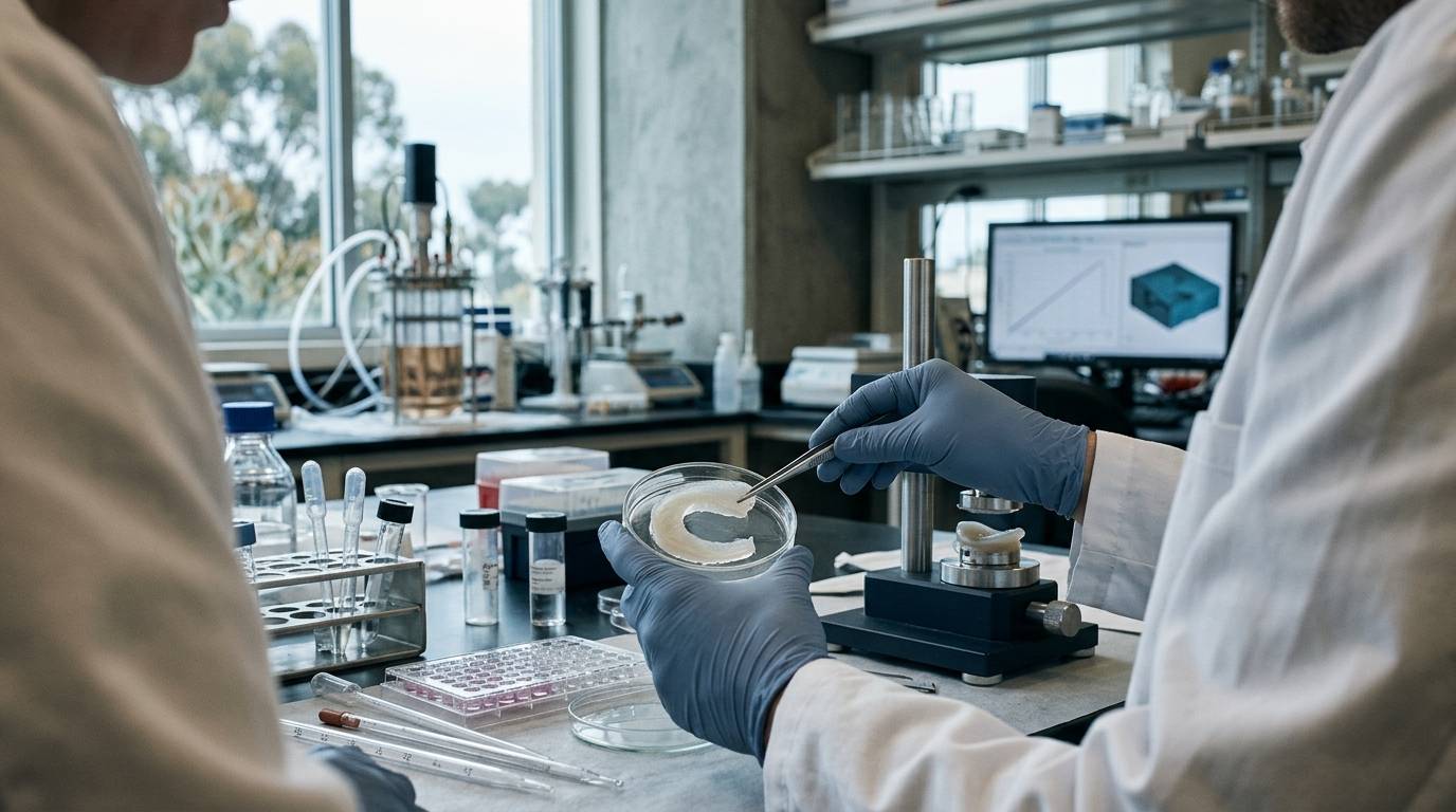
A research team places an experimental matrix into an articular defect, expecting rapid host cell migration. Weeks later, the defect remains largely vacant because joint tissues lack direct capillary networks. This scenario illustrates why investigating joint tissue repair requires an appreciation of structural constraints, specialized experimental models, and clear analytical readouts.
This guide outlines the unique physical and cellular properties of articular cartilage and meniscus tissue. It examines the baseline evidence supporting structural repair concepts and details how researchers assess repair outcomes in preclinical settings.
Structural constraints in joint tissue repair
Articular cartilage and the fibrocartilaginous meniscus possess minimal endogenous capacity for self-repair. Understanding why repair protocols often fail requires examining native tissue composition.
According to a review of human articular cartilage physiology, the tissue is avascular, aneural, and sparsely populated with cells. Because it lacks blood vessels, circulating progenitor cells and systemic nutrients cannot readily migrate to defect sites. The absence of a neural network also means early microtrauma generates no immediate local pain signals. This structural arrangement leaves damaged tissue with a severely restricted response to mechanical injury.
The same physiological review notes that articular cartilage consists of an extracellular matrix dominated by water, proteoglycans, collagen, and other functional proteins. These structural components interact to create a dense, highly pressurized fluid-solid matrix. This intricate architecture governs the tissue's mechanical behavior under compressive, tensile, and shear loads. When trauma disrupts this network, re-establishing identical matrix composition and mechanical resilience presents a major experimental hurdle.
Evaluating preclinical evidence levels
When designing studies, researchers must distinguish between theoretical physiological reviews and empirical repair trials. Much of the foundational literature describing matrix physiology reflects descriptive reviews of native human tissue. These reviews summarize structural traits but do not demonstrate the efficacy of specific therapeutic interventions.
Investigating tissue restoration requires controlled experimental studies. Preclinical studies typically evaluate specific scaffolds, cell therapies, or bioactive molecules within isolated laboratory models or animal joint systems. Researchers can consult mechanistic pathway resources to identify where preliminary cellular responses diverge from whole-joint biomechanics.
Reviewing published literature also requires distinguishing between in vitro binding observations and actual tissue remodeling. When analyzing published literature, interpreting preclinical peptide research requires cautious evaluation to avoid mistaking early bench assays for established clinical outcomes.
Animal models in joint repair studies
Preclinical joint repair investigations rely on animal systems to replicate physiological joint loading, synovial fluid dynamics, and complex geometry. Large-animal models remain standard for meniscus and chondral research because small rodents cannot mimic human-scale joint kinematics.
A systematic review of large-animal meniscus repair literature examined studies utilizing species such as sheep, pigs, dogs, and goats. These models enable researchers to simulate partial or total meniscal tears and test prospective repair scaffolds. However, researchers must account for differences in biomechanics, gait, cartilage thickness, and intrinsic cellularity across these species. Findings gathered from sheep or canine knees cannot be generalized directly to human clinical applications.
Common outcome measures in repair research
Evaluating repair tissue requires multiple independent readouts to confirm structural integrity, cellular attachment, and durability. Relying on a single metric frequently introduces bias into repair assessments.
According to the systematic review of large-animal meniscus repair studies, researchers employ several established outcome categories:
Histological analysis
Histology was the most frequent outcome metric, conducted in 90 percent of the reviewed studies. Histological staining identifies cellular morphology, cell density, and extracellular matrix organization. Specialized stains highlight the presence of proteoglycans and collagen fibers, revealing whether new tissue resembles native fibrocartilage or mechanically inferior fibrous scar tissue.
Macroscopic assessment
Macroscopic scoring occurred in 85 percent of the analyzed investigations. This method involves visual grading of defect filling, tissue surface smoothness, color, and margin integration under gross examination. While useful for initial screening, macroscopic assessment alone cannot determine cellular architecture or load-bearing capability.
Healing and integration parameters
Integration analysis was performed in 83 percent of the reviewed studies. These evaluations specifically measure the physical bond between the native host tissue and the implanted scaffold or reparative tissue. Incomplete peripheral integration frequently leads to mechanical failure under cyclic joint movement.
Secondary structural and functional metrics
The systematic review also identified other critical outcome parameters across large-animal studies:
- Biomechanical testing to measure compressive stiffness, tensile strength, and load redistribution across the joint surface.
- Biochemical and molecular assays to quantify specific collagen types, total water content, and sulfated glycosaminoglycan concentrations.
- Non-invasive imaging such as magnetic resonance imaging (MRI) to track tissue morphology and defect filling over longitudinal timepoints.
- Second-look arthroscopy to monitor tissue survival and surface integrity without terminating the animal model.
Understanding how analytical measurements differ from biological performance is critical. Researchers can inspect guides on separating binding affinity from functional biological effects to avoid misinterpreting physical interaction readouts as tissue regeneration.
Limitations of current experimental readouts
While preclinical animal literature provides valuable insight into repair mechanics, significant experimental uncertainties remain. One major limitation is the frequent disconnect between histological appearance and long-term joint function. An implant may achieve impressive macroscopic filling and tissue integration while failing to replicate the native mechanical behavior required for true joint protection.
Furthermore, variability in outcome reporting complicates cross-study comparisons. Because experimental methodologies vary across laboratories, pooling quantitative performance metrics across different large-animal designs remains difficult. Preclinical studies also typically evaluate outcomes across relatively short time horizons, leaving long-term matrix durability and secondary osteoarthritic changes poorly characterized.
Analytical controls and laboratory testing
Tissue engineering and regenerative research protocols frequently involve exogenous bioactive molecules, recombinant proteins, or peptide sequences designed to stimulate cell proliferation. Using research-use-only materials demands strict laboratory validation before beginning in vitro or in vivo experiments.
Verifying compound purity, chemical identity, and peptide concentration requires reliable analytical techniques such as high-performance liquid chromatography and mass spectrometry. A Certificate of Analysis (COA) confirms the analytical profile and purity of the specific tested sample only. A COA does not predict in vivo biological activity, tissue integration, or systemic safety. Researchers should consult laboratory quality testing standards to understand the precise capabilities and limitations of analytical test records.
When to revisit this resource
Revisit this resource when designing preclinical joint defect protocols, selecting outcome measures for meniscus research, or evaluating structural readouts in published repair literature. Rigorous tissue repair investigations depend on recognizing the natural limits of avascular joint structures and pairing structural findings with verified functional testing.
Sources
Introducing / CaliLean Field Notes
A little more
perspective.
An editorial letter for curious minds. Thoughtful reading, questions worth keeping and the sources behind them.
Explore Field Notes Join the list for the first edition

