
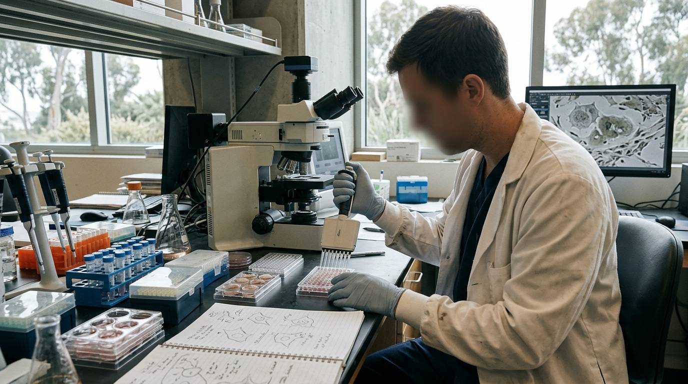
Many researchers search for whether cellular senescence helps or hinders acute tissue repair. The common assumption treats all senescent cells as damage that must be removed. This guide examines the experimental evidence behind cellular senescence in cutaneous wound models, explaining the specific mechanisms and laboratory methods used to evaluate these pathways.
Scope of Senescence in Tissue Repair
Senescence refers to a state of stable cell cycle arrest triggered by cellular stress or programmed physiological cues. In regenerative biology, researchers study how temporary senescent populations interact with neighboring cells. Investigating these dynamics requires precise experimental models rather than broad assumptions about cellular aging. For more context on tissue models, see our recovery and repair research resources.
Wound healing requires coordinated signals across multiple cell types. While long-term accumulation of senescent cells can cause chronic dysfunction, transient senescence serves distinct functional roles. Experimental data show that certain senescent cells provide essential paracrine signals during acute matrix reconstruction.
Evidence Levels Across Cutaneous Models
Most mechanistic understanding of senescence in wound repair originates from genetically modified rodent models. Evidence from cell cultures and animal models should not be conflated with controlled human trials. To evaluate published studies critically, researchers often consult guides on how to read scientific research.
Currently, the direct causal role of senescent cells in wound closure is supported by animal experiments. Human data remain primarily observational, relying on tissue biopsies and marker staining. Controlled interventional human data targeting senescent cell ablation during acute cutaneous injury remain scarce.
Transgenic Animal Study Models
To examine the functional role of senescence in live tissue, researchers use specialized mouse strains. A prominent model is the p16-3MR transgenic mouse. In this model, the promoter for p16INK4a drives a functional cassette that allows tracking and selective clearance of p16-expressing cells.
Researchers administered ganciclovir to p16-3MR mice subjected to full-thickness dorsal punch biopsies. This method allowed the targeted death of p16-positive cells specifically during the healing phase. By tracking the wounds, investigators observed the exact phenotypic consequences of cell clearance.
Cell identification within these cutaneous wound margins revealed specific cell lineages entering senescence. The primary cells expressing senescent phenotypes were localized fibroblasts and vascular endothelial cells. Tracking these distinct lineages prevented overgeneralized conclusions across mixed cell types.
Molecular Mechanisms and Paracrine Pathways
Senescent cells modify their local microenvironment through active secretomes. In acute injury models, senescent fibroblasts and endothelial cells produce platelet-derived growth factor subunit A, also designated as PDGF-A. For a broader analysis of signaling networks, explore our review of molecular mechanisms and pathways.
This senescent-derived PDGF-A, forming homodimers known as PDGF-AA, acts directly on local target cells. It stimulates adjacent fibroblasts to undergo myofibroblast differentiation. Myofibroblasts are critical effector cells that generate contractile forces and synthesize provisional extracellular matrix.
When senescent cells produce PDGF-AA, wound contraction progresses normally. The presence of these transient populations helps organize the local tissue architecture. Thus, early senescence provides an essential signaling niche rather than simple cellular damage.
Findings from Targeted Depletion Studies
Direct experimental ablation of p16-positive cells produced measurable functional changes in cutaneous wounds. In the 2014 p16-3MR mouse study reviewed in Frontiers in Immunology, clearing senescent cells significantly delayed early wound closure.
The primary experimental findings demonstrated several specific outcomes:
- Full-thickness skin punch biopsies induced p16-positive senescent cells at the wound edge.
- Senescent fibroblasts and endothelial cells secreted high levels of PDGF-A.
- Secreted PDGF-AA triggered the activation of fibroblasts into functional myofibroblasts.
- Targeted elimination of these p16-positive cells retarded normal wound closure.
These observations confirm that senescent cells can execute beneficial, programmed functions in acute healing. Eliminating them indiscriminately can disrupt necessary signaling sequences.
Methodological Limitations and Experimental Uncertainties
The p16-3MR model depends on ganciclovir-mediated suicide gene activation. Off-target cellular stress or incomplete ablation can complicate data interpretation. Furthermore, rodents heal primarily through panniculus carnosus contraction, whereas human cutaneous repair relies heavily on re-epithelialization and granulation.
Another key unknown is the threshold between transient beneficial senescence and chronic maladaptive senescence. In chronic non-healing ulcers, prolonged secretome release may impair cellular migration. Researchers cannot extrapolate findings from acute, clean incisions directly to chronic human wounds.
Marker ambiguity also creates methodological hurdles in laboratory research. Markers such as p16INK4a and senescence-associated beta-galactosidase are not wholly specific to irreversible arrest. Overreliance on single markers can misidentify reversible quiescence or terminal differentiation as cellular senescence.
Analytical Considerations for Laboratory Reagents
Evaluating senescence pathways in vitro requires rigorous analytical verification of research reagents. When testing synthetic peptides or growth factor ligands in culture, researchers verify chemical identity through proper analytical methods. High-performance liquid chromatography and mass spectrometry establish purity and molecular mass for experimental lots.
Researchers reviewing quality documentation should consult standards on reading a Certificate of Analysis. A Certificate of Analysis confirms only that a specific tested sample met analytical parameters. It cannot predict biological behavior in complex cellular assays or validate clinical utility.
Protocol Evaluation Checklist
Researchers designing experiments to investigate senescence in tissue repair can apply this verification checklist:
- Validate multiple senescence markers: Combine p16INK4a detection with p21 expression and senescence-associated beta-galactosidase assays rather than using a single marker.
- Characterize the secretome: Quantify specific ligands such as PDGF-A in conditioned medium before attributing downstream phenotypic changes to cell arrest.
- Distinguish model mechanics: Account for contraction versus re-epithelialization when interpreting murine wound closure timelines relative to human tissue repair.
- Verify research materials: Confirm lot-specific purity and molecular weight using analytical mass spectrometry and chromatography documentation before initiating assays.
Sources
Introducing / CaliLean Field Notes
A little more
perspective.
An editorial letter for curious minds. Thoughtful reading, questions worth keeping and the sources behind them.
Explore Field Notes Join the list for the first edition

