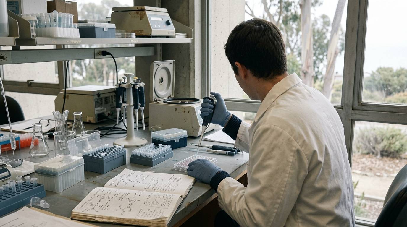
A researcher measures peptide clearance in rodent plasma, observes rapid cleavage, and assumes the same rate applies across mammalian systems. Later assays in non-human primate or human matrices reveal unexpected differences in enzymatic stability and metabolite proportions.
Peptide degradation pathways, clearance rates, and secondary metabolite profiles frequently diverge across experimental test systems. This guide reviews how laboratory models differ during peptide metabolism experiments. It details empirical findings across species and outlines reporting practices required to evaluate model suitability.
Scope of Model Comparisons in Peptide Science
This resource addresses how peptide clearance and enzymatic degradation vary across experimental test systems. Evaluating research models requires clear definitions of the peptide under review, the biological matrix examined, and the specific enzymes responsible for cleavage. In peptide research, biological activity depends heavily on structural preservation. When a peptide enters a biological fluid, host peptidases can cleave terminal residues or internal bonds almost immediately.
Assessing model suitability requires researchers to avoid treating mammalian systems as interchangeable units. Metabolic speed, dominant cleavage sites, and downstream fragments change when moving from cell-free matrices to animal models. Understanding these shifts helps laboratories interpret preclinical literature without generating unsupported translational claims.
Evaluating the Levels of Evidence
Metabolic peptide studies rely on distinct tiers of empirical data. Laboratory investigations typically begin with in vitro plasma incubations. These models isolate fluid-phase enzymatic degradation from organ clearance. They allow tight control over temperature and substrate concentrations. However, in vitro assays omit tissue-bound peptidases, renal filtration, and hepatic extraction.
In vivo animal models introduce physiological clearance mechanics, whole-body perfusion, and multi-organ processing. These models measure how intact animals process parent peptides and cleared fragments over time. Controlled human data remain the reference point for clinical pharmacology. Yet, in vitro animal findings cannot substitute for human clinical trials. Each tier reveals unique parameters of peptide stability, which makes cross-model comparisons essential. Readers tracking study design methodology can reference our guide on peptide research evidence levels.
Model Systems and Interspecies Divergence
Metabolic observations depend strictly on the biological model employed. Variations between rodent species, non-human primates, and human plasma reflect differences in circulating peptidase concentrations and substrate specificity.
In Vitro Plasma Degradation Models
In vitro plasma assays allow researchers to compare initial clearance kinetics across multiple species under identical conditions. A direct example comes from an in vitro comparative study examining dynorphin A1 to 13 degradation across multiple mammalian matrices. The investigators tested plasma from monkeys, rabbits, rats, and guinea pigs, and compared these profiles against previously reported human data.
The study demonstrated that even when general degradation pathways are conserved, baseline kinetics shift substantially across species. The authors observed rapid processing across all tested matrices. Half-lives ranged from 0.5 minutes in guinea-pig plasma to 1.1 minutes in monkey plasma. Furthermore, downstream processing diverged after the initial cleavage event.
Rodent In Vivo Experimental Systems
In vivo experiments introduce systemic clearance pathways that in vitro fluids lack entirely. A study in C57BL/6JRj mice evaluated glucagon-like peptide-1 (GLP-1) dynamics to determine how physiological stimuli influence peptide handling. Researchers administered oral glucose, a mixed meal, or water to 256 mice, collecting blood at multiple time intervals between 2 and 90 minutes.
To track elimination directly, investigators also administered the primary metabolite, GLP-1(9-36)NH2, intravenously to 160 mice. This experimental setup showed that intact physiological systems process peptides through layered enzymatic cascades. When evaluating in vivo studies, researchers should consult frameworks on how to interpret peptide study data to prevent conflating local degradation with total biological clearance.
Cleavage Pathways and Peptidase Mechanisms
Peptides undergo breakdown through precise enzymatic actions, including aminopeptidases, carboxypeptidases, and neutral endopeptidases. The exact enzyme driving degradation can vary across species and experimental models.
In the dynorphin A1 to 13 comparative investigation, researchers utilized enzyme inhibition alongside kinetic analysis to identify cleavage mechanisms. Both approaches yielded aligned results. The primary degradation pathway was broadly similar across the animal matrices, generating dynorphin A1 to 12 as the major initial metabolite. However, the exact proportion of this metabolite varied by species, representing 71% of breakdown products in rabbit plasma compared to 88% in guinea-pig plasma. Subsequent breakdown of dynorphin A1 to 12 displayed even greater heterogeneity across monkey, rabbit, rat, and guinea-pig plasma.
In the mouse
GLP-1 investigation, researchers identified rapid elimination of the peptide fragment within minutes. The authors found evidence implicating neutral endopeptidase (neprilysin or NEP 24.11) as a primary driver of degradation in mice. This endoproteolytic action cleaves the peptide beyond the initial dipeptidyl peptidase-4 (DPP-4) site. This finding shows that downstream cleavage can obscure the parent compound. It also demonstrates how tissue or circulating endopeptidases alter the half-life of intermediate metabolites.
Analytical Findings Across Test Systems
The empirical literature confirms that peptide recovery and kinetic measurements depend heavily on species selection and analytical tooling.
First, half-life values cannot be directly translated across species. The in vitro dynorphin study confirmed that monkey plasma retained the peptide more than twice as long as guinea-pig plasma. Intermediate metabolite generation also followed distinct patterns. The higher proportion of dynorphin A1 to 12 in guinea pigs indicates species-specific enzymatic affinity or differential inhibitor sensitivity.
Second, in vivo measurement accuracy depends on assay architecture and cleavage site awareness. In the mouse GLP-1 study, researchers found that measured GLP-1 profiles differed substantially depending on the assay platform used. Conventional sandwich enzyme-linked immunosorbent assays (ELISAs) were inadequate for monitoring murine peptide concentrations. This failure occurred because neprilysin-mediated endoproteolytic cleavage cleaved the epitope target generated by DPP-4. Accurately tracking GLP-1 secretion in mice therefore required specialized assays capable of capturing NEP 24.11 metabolites.
These findings show that detection methods must match the exact proteolytic pathways of the chosen animal model. When assays lack sensitivity to specific fragments, data may reflect detection failure rather than actual biological clearance. Exploring the relationship between binding affinity and biological effect can clarify how fragment generation influences overall experimental interpretations.
Methodological Limitations and Reporting Standards
Translating peptide metabolism data between experimental systems involves notable analytical hurdles. In vitro plasma experiments exclude vital clearance routes, such as renal filtration, bile excretion, and membrane-bound enzymatic processing in organs like the lungs or kidneys. Consequently, an in vitro plasma half-life represents an isolated enzymatic rate, not total systemic clearance.
Analytical cross-reactivity creates another layer of experimental uncertainty. If an assay cannot distinguish between a full-length peptide and truncated fragments, calculated concentration curves will be incorrect. The GLP-1 research shows that failing to account for secondary endoproteolytic cleavage produces invalid clearance profiles in mouse models.
To resolve inconsistency in preclinical literature, researchers rely on established methodology frameworks rather than assuming model uniformity. The ARRIVE 2.0 guidelines provide an official reporting framework for animal experiments. Importantly, ARRIVE 2.0 is a standardized reporting guideline, not an experimental paper demonstrating biological strain differences. It details necessary disclosures for animal sex, strain, housing, and analytical verification, ensuring independent laboratories can replicate reported conditions.
Further reading in our research methods library provides context on structured experimental reporting and literature evaluation.
When to Revisit This Resource
Revisit this resource when designing interspecies comparative protocols, selecting immunoassay reagents for rodent studies, or auditing peptide clearance literature for translational consistency.
Careful selection of biological matrices and analytical assays ensures peptide metabolism research remains rigorous, reproducible, and transparent.
Sources
Introducing / CaliLean Field Notes
A little more
perspective.
An editorial letter for curious minds. Thoughtful reading, questions worth keeping and the sources behind them.
Explore Field Notes Join the list for the first edition