
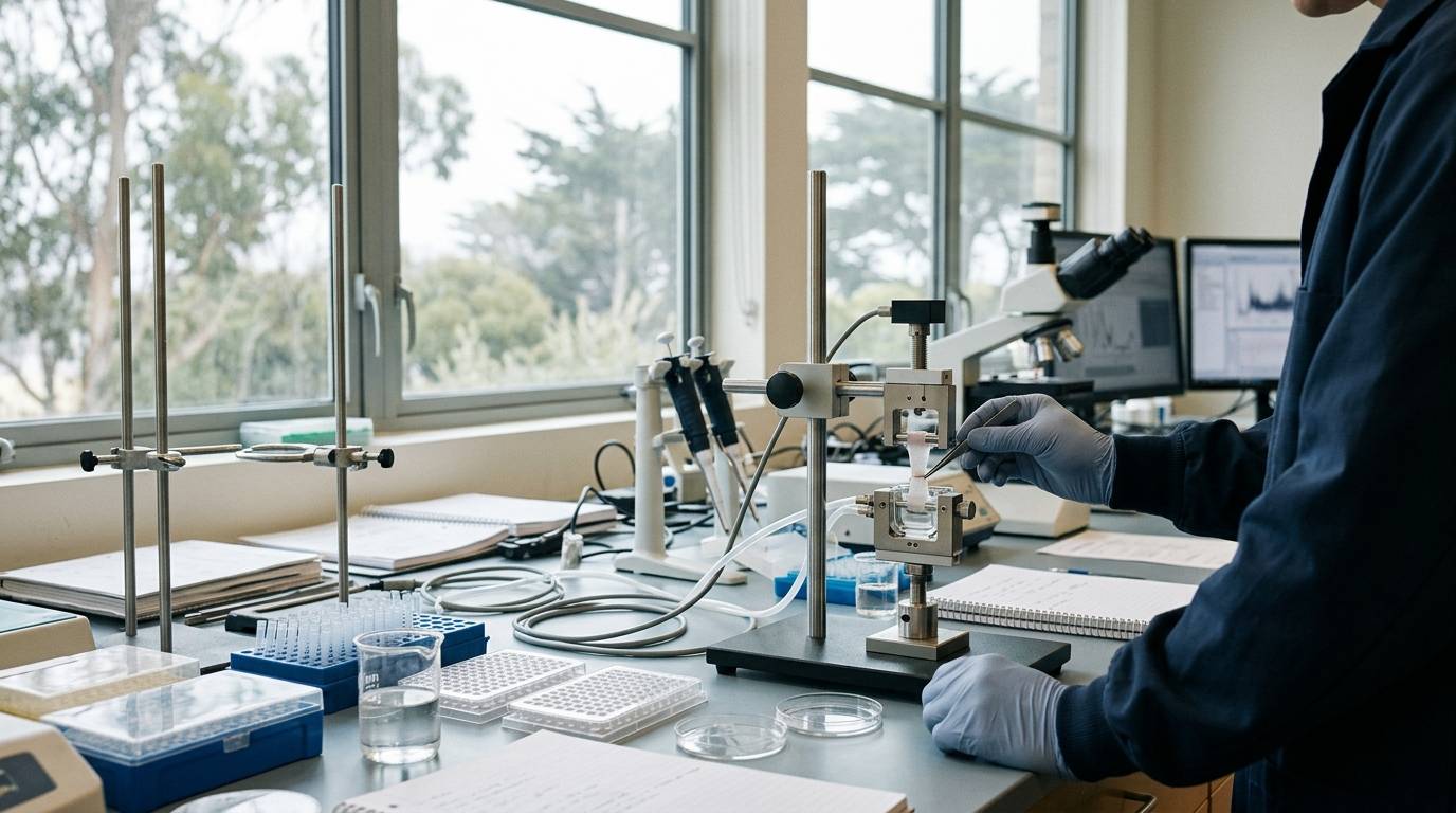
Why do tissue repair studies frequently report complete structural healing or positive gene expression, yet fail to demonstrate functional mechanical integrity? Researchers often encounter this exact discrepancy when evaluating experimental recovery data. This resource outlines how to select endpoints that distinguish molecular activity, structural continuity, and functional load-bearing capacity.
Endpoint Hierarchy in Experimental Repair Models
Experimental repair evaluations require clear boundaries between distinct biological tiers. A molecular signal indicates that a cellular pathway responded to an intervention. However, an altered transcription profile does not guarantee tissue reorganization. Similarly, structural bridging under macroscopic observation does not demonstrate that a repaired structure can withstand mechanical load.
A rigorous tissue repair study evaluates three complementary categories of endpoints:
- Molecular markers, such as transcript levels and inflammatory mediators.
- Structural markers, such as histology, cellular infiltration, and imaging.
- Functional markers, including failure force, stiffness, and tensile strength.
Relying on a single metric risks false conclusions about recovery. To understand how molecular interactions translate into phenotypic changes, researchers must recognize the distance between binding and functional outcomes. Defining endpoints across all three tiers ensures that chemical observations align with physical integrity.
Methodological Context and Evidence Levels
The primary literature underlying these endpoint relationships relies predominantly on controlled animal models. Preclinical studies isolate specific genetic or physical variables within standardized laboratory environments. These animal experiments provide essential mechanistic data, but they carry distinct limitations that prevent direct extrapolation to human clinical outcomes.
Evaluating experimental design requires distinguishing preclinical models from clinical observation. As outlined in our guide to interpreting preclinical study designs, in vivo animal models demonstrate proof of mechanism rather than therapeutic efficacy. Controlled rodent investigations clarify how tissues reorganize under specific, regulated injuries. They do not eliminate the necessity for standardized validation when moving across species.
Characterization of Validated Animal Models
Experimental validation of repair endpoints requires precise surgical and genetic models. Two specific mouse models illustrate the divergence between visual healing and mechanical restoration.
Murine Dermal Incision Model
One validated model evaluates incisional dermal healing in genetically altered mice. This setup compares wild-type mice against prostaglandin H synthase-1 (pghs-1) null mice and prostaglandin H synthase-2 (pghs-2) null mice. Investigators execute standardized surgical incisions to assess dermal repair across three phases: inflammatory, proliferative, and remodeling phases.
The analytical protocol for this model tracks wound tensile strength alongside transcript levels of pghs-1 and pghs-2 at the injury site. Additionally, histological sections quantify cellular migration, inflammatory infiltration, and matrix deposition. This design allows investigators to isolate the influence of specific enzymes on structural repair versus functional mechanical performance.
Murine Rotator Cuff Repair Model
A second standardized model assesses tendon-to-bone integration using sixty male C57BL/6 mice. In this procedure, surgeons perform a transection of the rotator cuff tendon followed by open microsurgical repair. A modified Kessler suturing technique secures the tendon prior to complete detachment.
The sutures pass through two intersecting bone tunnels placed at the anatomic insertion footprint. Investigators evaluate the repair at two and four weeks post-surgery. The testing matrix includes micro-computed tomography (micro-CT), histological grading, biomechanical failure testing, and targeted gene expression profiling.
Structural Continuity Versus Functional Failure Force
The audited animal literature demonstrates that tissue continuity often masks functional deficits. When investigators evaluate repair using visual inspection alone, critical mechanical weaknesses remain undetectable.
In the dermal incision study, wounds across wild-type, pghs-1 null, and pghs-2 null mice appeared macroscopically indistinguishable during gross inspection. Visual assessment suggested identical closure. However, biomechanical tensile testing revealed significant functional impairments at day 12 after incision. The healed wounds in pghs-1 null mice reached only 75% of wild-type tensile strength. Wounds in pghs-2 null mice reached only 70% of wild-type strength.
- Dermal Incision Tensile Strength at Day 12
- Wild-type: 100% of baseline strength
- pghs-1 null: 75% of wild-type strength
- pghs-2 null: 70% of wild-type strength
A similar discordance occurs in musculoskeletal models. In the C57BL/6 rotator cuff repair experiment, histological sections at two and four weeks revealed complete reattachment of the tendon to the bone footprint. High-resolution imaging documented no observable physical gap between the tissues. Instead, the interface filled with fibrovascular scar tissue, mirroring the repair patterns observed in larger animal species.
- Rotator Cuff Biomechanical Failure Force
- Intact tendon: 100% failure force
- 2-week repair: 36% of intact tendon force
- 4-week repair: 75% of intact tendon force
Despite the complete reattachment seen on histology, biomechanical testing showed that failure force remained depressed. At two weeks, failure force reached only 36% of the value of an intact, uninjured tendon. By four weeks, failure force recovered to 75% of intact tendon values. Gene expression analysis demonstrated molecular alterations consistent with gradual healing across the four-week timeline. However, gene expression alone did not indicate when full mechanical competence was achieved.
Analytical Limitations and Preclinical Gaps
Relying on isolated biomarker panels or structural imaging creates notable experimental risks. Fibrovascular tissue bridges physical gaps rapidly, creating the visual appearance of union in histological cross-sections. Yet this disorganized scar tissue possesses inferior collagen organization, which compromises peak failure load.
Biomechanical endpoints are technically demanding and require destructive testing methods. Because a specimen cannot undergo tensile testing and subsequent intact histology simultaneously, studies require higher animal numbers across parallel cohorts. Micro-CT provides structural density values for surrounding mineralized bone, but it cannot evaluate soft-tissue shear strength directly.
Furthermore, transcript abundance does not mirror functional protein assembly. Messenger RNA expression indicates active transcription at a specific time point. It does not measure mature collagen cross-linking, enzymatic degradation rates, or mechanical resistance. Researchers utilizing rigorous research methods must combine histological continuity, transcript levels, and mechanical metrics to construct an accurate assessment of tissue healing.
Checklist for Designing Repair Study Endpoints
Apply this operational checklist when constructing preclinical tissue-repair protocols:
- Define the primary functional threshold: Determine the specific mechanical parameter, such as failure force, stiffness, or tensile strength, required to establish functional recovery.
- Select non-destructive structural metrics: Integrate high-resolution imaging, such as micro-CT, to evaluate mineral and architectural features before destructive biomechanical tests.
- Map time courses across biological phases: Establish collection windows that capture inflammatory, proliferative, and remodeling phases rather than a single end point.
- Pair gene panels with histology: Align transcriptional markers with localized tissue sections to confirm that mRNA signals reflect actual protein and cellular organization.
- Account for scar tissue formation: Screen histological samples for disorganized fibrovascular tissue that provides physical closure without native tensile capacity.
Sources
Introducing / CaliLean Field Notes
A little more
perspective.
An editorial letter for curious minds. Thoughtful reading, questions worth keeping and the sources behind them.
Explore Field Notes Join the list for the first edition

