
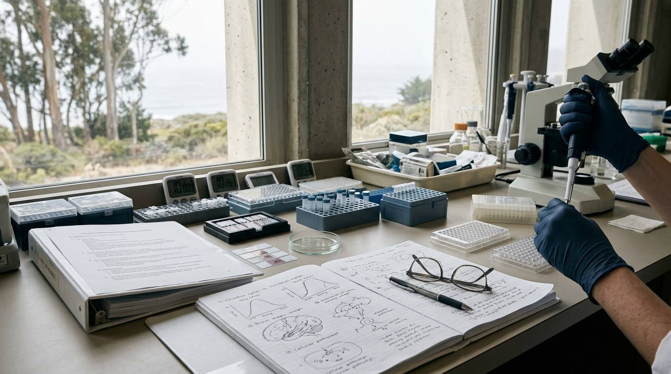
Standard pharmacological protocols assume that biological tissue presents a static target across the twenty-four-hour day. In central nervous system pharmacology, researchers often treat the time of compound administration as an operational convenience rather than an active independent variable. This assumption introduces systematic error into preclinical and translational neuroscience. A neural substrate sampled at dawn does not possess the same transcriptional landscape, receptor density, or synaptic tone as the identical substrate sampled at dusk.
When an exogenous peptide or synthetic analogue is administered in vivo, the observed phenotype reflects more than compound pharmacology. The measured biological response represents an interaction among molecule exposure, internal circadian phase, accumulated homeostatic sleep pressure, and acute environmental cues. Failing to isolate these variables produces irreproducible results, contradictory potency estimates, and obscured mechanistic pathways.
Evaluating peptide signaling in the brain requires experimental designs that treat biological time as a primary structural factor. This guide outlines the conceptual models, empirical controls, and analytical frameworks required to isolate peptide-driven effects from the endogenous rhythms of the central nervous system.
Establish Biological Time as an Experimental Variable
A compound does not act upon an inert baseline. In the brain, intracellular cascades, membrane potentials, and neuropeptide receptor expressions oscillate along predictable twenty-four-hour trajectories. If an investigator administers a test compound at nine in the morning and observes altered locomotor activity, attributing that outcome purely to peptide efficacy is a methodological error. The response might occur because the target receptor peaks at that specific hour. Conversely, the compound might simply shift the phase of an underlying behavioral oscillator.
To address this challenge, researchers must define their experimental question with mathematical precision. Investigators should refer to guidelines on peptide research evidence levels to distinguish between preliminary observational findings and robust causal models.
Before designing an experiment, researchers must determine whether they are measuring a static treatment effect or a dynamic interaction. A baseline study might ask if a compound changes an endpoint at one single phase. A more rigorous chronobiological study asks whether the magnitude or direction of that response varies as a function of internal phase.
- Estimand Types in Peptide Chronobiology
- Phase-Specific Treatment Effect: Quantifying response at a fixed, predefined biological time.
- Endogenous Oscillation: Mapping baseline rhythmicity in vehicle-treated control cohorts across twenty-four hours.
- Treatment-by-Phase Interaction: Evaluating whether compound efficacy depends on internal biological timing.
- Phase-Shifting Capacity: Testing whether an intervention advances or delays the peak timing of an endogenous rhythm.
- Waveform Modification: Determining whether treatment alters rhythm amplitude, baseline mean, or harmonic complexity.
Treating biological time as an experimental variable transforms how protocols are scheduled. Assays cannot simply occur when laboratory personnel arrive in the morning. Experimental time points must be distributed systematically across the internal biological cycle of the target organism.
Differentiate Circadian Oscillations from Sleep Homeostasis
Investigating brain peptides that modulate arousal, cognition, or vigilance requires separating two distinct physiological drives. Investigators rely on the classic two-process model of sleep regulation to distinguish these forces. The two-process model separates sleep propensity into Process S and Process C. These systems run simultaneously, but their regulatory mechanisms and molecular drivers are fundamentally distinct.
Process S represents homeostatic sleep pressure. It builds continuously during sustained wakefulness and dissipates exponentially during slow-wave sleep. If an animal remains awake across its active phase, Process S rises steadily. In contrast, Process C represents the endogenous circadian rhythm of sleep propensity. Process C oscillates across approximately twenty-four hours under the control of an internal pacemaker, completely independent of whether the organism is asleep or awake.
- Core Regulatory Drivers
- Process S (Homeostatic Drive): Wake-dependent accumulation of somnogenic pressure; dissipates during rest.
- Process C (Circadian Drive): Autonomous oscillatory gating; driven by the central suprachiasmatic pacemaker.
- Observed Arousal State: The physiological summation of Process S and Process C interactions.
When an experimental design notes that a subject was tested in the early morning, that notation tells an incomplete story. Two subjects tested at the identical hour can occupy radically divergent physiological states. One subject may possess high homeostatic sleep pressure due to prior sleep deprivation, while another carries low sleep pressure after full rest. Yet both subjects may share an identical internal circadian phase.
Researchers must track and standardize prior wakefulness alongside biological timing. If an animal model receives a neuropeptide after extended wakefulness, an investigator cannot cleanly determine whether the compound modulated circadian pathways or interacted with elevated homeostatic sleep pressure. Both variables must be quantified, documented, and held constant across treatment arms. For broader context on structuring these protocols, consult foundational research methods resources.
Environmental cues further complicate this balance through a process known as masking. Masking occurs when an acute external stimulus directly alters a physiological endpoint, bypassing the central circadian pacemaker entirely. Bright light acutely suppresses locomotor activity in nocturnal rodents, while promoting alertness in diurnal humans. This direct environmental suppression can easily be mistaken for an endogenous circadian shift or a compound-mediated effect.
To prevent masking from confounding experimental conclusions, investigators must separate environmental time from biological time. Standard experimental protocols rely on Zeitgeber Time, abbreviated as ZT. In a standard twelve-hour light and twelve-hour dark environment, ZT0 marks the precise onset of light, while ZT12 marks lights-off. Zeitgeber Time directly indexes the subject to an external lighting cycle.
- Time Conventions in Chronobiological Research
- Zeitgeber Time (ZT): Time relative to an external environmental cycle (ZT0 lights-on, ZT12 lights-off).
- Circadian Time (CT): Time under constant environmental conditions (CT0 subjective dawn, CT12 subjective dusk).
- Clock Time: Local civil wall-clock time; carries zero biological meaning without environmental context.
When research questions require removing environmental zeitgebers entirely, investigators transition subjects into constant darkness or constant dim light. Under these constant conditions, Zeitgeber Time becomes invalid. Researchers must transition to Circadian Time, abbreviated as CT. CT0 represents the onset of the organism's subjective day, while CT12 indicates the onset of the subjective night.
Equating clock time, ZT, and CT is a widespread source of experimental error. A report that describes animal procedures using wall-clock hours without defining the light cycle prevents independent replication. Researchers must explicitly declare their timing conventions, baseline photoperiods, and light-phase parameters in every published methodology.
Classify Brain Peptide Signaling Across Network and Output Circuits
Neuropeptides in the mammalian central nervous system do not occupy a uniform tier within the circadian hierarchy. When planning an investigation, researchers must categorize candidate peptides based on their functional position within oscillatory neural networks. Peptides generally operate as core network synchronizers, downstream arousal modulators, or phase-dependent physiological outputs.
- Functional Tiers of Circadian Neuropeptides
- Pacemaker Network Synchronizers: Peptides maintaining intercellular synchrony within the suprachiasmatic nucleus (e.g. VIP, AVP).
- Arousal and Vigilance Regulators: Hypothalamic peptides governing sleep-wake state transitions (e.g. Orexin, MCH).
- Phase-Dependent Output Signals: Neuroendocrine peptides whose release and receptor sensitivity fluctuate downstream.
The master mammalian pacemaker resides within the suprachiasmatic nucleus of the anterior hypothalamus. Individual neurons within this nucleus possess autonomous molecular clocks driven by transcriptional-translational feedback loops. However, isolated cellular oscillators exhibit divergent periods and drift without intercellular coordination. Coherent twenty-four-hour tissue rhythmicity requires robust peptidergic coupling across the neural network.
Vasoactive intestinal peptide, commonly designated VIP, serves as a primary synchronizing agent within the central pacemaker. Expressed densely in the ventral suprachiasmatic nucleus, VIP acts upon VPAC2 receptors expressed across adjacent pacemaker neurons. VIP-VPAC2 signaling modulates intracellular cyclic adenosine monophosphate, alters membrane excitability, and regulates immediate-early gene expression. This peptidergic cascade synchronizes disparate cellular pacemakers into a unified tissue-level rhythm that entrains to environmental light cues.
Investigators must recognize that VIP signaling does not operate according to simple linear dose-response kinetics. Cell-culture and tissue-explant studies demonstrate that intermediate levels of VIP-VPAC2 signaling maximize network synchrony among pacemaker neurons. Reducing VIP tone destabilizes cellular phase coherence. Paradoxically, saturating the network with excessive VIP concentrations also degrades synchrony.
- Nonlinear Dynamics of VIP-VPAC2 Signaling
- Low VIP Signaling: Uncouples individual pacemakers; leads to dispersed phase distribution across the network.
- Intermediate VIP Signaling: Optimizes intercellular coupling; establishes robust, high-amplitude tissue rhythmicity.
- Excessive VIP Signaling: Desensitizes VPAC2 receptors; disrupts network synchronization and impairs rhythmic output.
Arginine vasopressin, or AVP, represents another core peptidergic network signal within the suprachiasmatic nucleus. AVP is expressed rhythmically within the dorsal shell of the nucleus, operating alongside gamma-aminobutyric acid to sculpt rhythmic inhibitory output. VIP, AVP, and GABA form a tightly integrated paracrine circuit. An intervention that targets one neuropeptide pathway alters the activity of the interconnected network.
Downstream from the central pacemaker lie the arousal networks of the lateral hypothalamus. Neurons producing orexin, also known as hypocretin, project broadly throughout the brain to sustain wakefulness and vigilance. Orexin neurons fire vigorously during active wake states and fall silent during non-rapid eye movement and rapid eye movement sleep. Prepro-orexin transcript levels, mature peptide abundance, and orexin receptor densities all exhibit pronounced twenty-four-hour rhythmicity under baseline conditions.
Recent research demonstrates that lateral hypothalamic signaling feeds back directly into the central pacemaker. Both orexin and melanin-concentrating hormone, or MCH, modulate the intrinsic circadian period of suprachiasmatic tissue in ex vivo preparations. Peptides traditionally categorized as simple downstream sleep regulators can actively remodel upstream pacemaker dynamics.
To design rigorous experiments, investigators must precisely define their biological endpoints. Tracking changes in steady-state peptide abundance does not demonstrate rhythmic release. Similarly, rhythmic neuropeptide release does not guarantee rhythmic receptor activation or downstream behavioral change. Each step in the signaling cascade requires independent validation through targeted assays. For specialized literature addressing central neuropeptide mechanisms, explore the brain, sleep, and cognitive research directory.
Separate Compound Dynamics from Phase-Dependent Receptor Sensitivity
When a candidate peptide produces divergent biological responses across the day, two distinct mechanisms may explain the variation. The difference could arise from chronopharmacokinetics, meaning the compound's absorption, distribution, metabolic clearance, or central penetration fluctuates over time. Alternatively, the difference could stem from chronopharmacodynamics, where tissue concentration remains constant but target receptor sensitivity oscillates.
Chronopharmacology requires researchers to measure internal exposure alongside physiological endpoints. If an investigator administers an intranasal or peripheral peptide and measures higher central activity at ZT6 than at ZT18, the pharmacokinetic profile must be mapped first. Blood-brain barrier transporter activity, local enzymatic degradation, and glymphatic clearance vary across the day.
- Mechanistic Drivers of Time-Dependent Pharmacology
- Chronopharmacokinetics: Phase-dependent variation in compound absorption, barrier penetration, and metabolic clearance.
- Chronopharmacodynamics: Temporal fluctuations in target receptor density, G-protein coupling, or downstream kinase activity.
- Clearance Oscillations: Glymphatic flow and enzymatic degradation rates that fluctuate across sleep-wake states.
If pharmacokinetic metrics show identical exposure profiles across different administration times, the variation must stem from chronopharmacodynamics. Receptor availability changes dynamically across the circadian cycle. Receptors internalize, recycle, and alter their coupling efficiency to downstream G-proteins and beta-arrestins based on circadian phase.
For example, VIP-induced intracellular calcium responses in suprachiasmatic neurons show profound temporal gating. In isolated neuronal populations, applying VIP during the subjective day significantly dampens intracellular calcium levels. Applying the identical concentration of VIP during the subjective night produces negligible calcium changes. The extracellular peptide concentration is identical, but the internal state of the target cell dictates the functional outcome.
Investigators can avoid conflating these mechanisms by employing clean multi-tiered protocols:
- Analytical Workflow for Disentangling Chronopharmacology
- Map Target Tissue Exposure: Measure compound concentration profiles in target brain structures across multiple phases.
- Characterize Receptor Expression: Quantify target receptor mRNA and membrane-bound protein levels at each time point.
- Assess Downstream Activation: Measure immediate intracellular intermediates (such as cAMP or phosphorylation cascades).
- Evaluate Phenotypic Output: Correlate biochemical signaling directly with behavioral or neurophysiological metrics.
Researchers can cross-reference foundational biochemical principles by reviewing peptide compound identity and sequence validation resources. Establishing molecular purity and sequence fidelity ensures that divergent signaling reflects endogenous biology rather than compound breakdown.
Structure Experimental Designs to Isolate Treatment-by-Phase Interactions
Standard parallel-group designs that test compounds at a single convenient time point are inadequate for chronobiological research. If an investigator compares a peptide-treated group to a vehicle group exclusively at ZT2, the study yields zero information about the circadian system. The observed difference might reverse entirely if measured twelve hours later.
To resolve time-dependent effects, researchers should deploy factorial designs that cross compound treatment with circadian phase. In a two-by-four factorial framework, subjects are assigned to either vehicle or peptide treatment and evaluated across four distinct Zeitgeber times, such as ZT0, ZT6, ZT12, and ZT18.
- Factorial Layout (Treatment x Phase)
- Group A1: Vehicle at ZT0 Group B1: Peptide at ZT0
- Group A2: Vehicle at ZT6 Group B2: Peptide at ZT6
- Group A3: Vehicle at ZT12 Group B3: Peptide at ZT12
- Group A4: Vehicle at ZT18 Group B4: Peptide at ZT18
This structural architecture permits the statistical model to estimate three distinct terms: the main effect of the peptide, the main effect of biological time, and the interaction term between treatment and time. A significant interaction term demonstrates that peptide efficacy depends strictly on internal phase.
When evaluating behavioral or cognitive endpoints, such as spatial memory or fear extinction, testing-time effects frequently confound training-time outcomes. An animal trained at ZT3 and tested at ZT15 may perform poorly not because memory consolidation failed, but because retrieval processes oscillate. To untangle these factors, investigators utilize counterbalanced crossover designs.
- Counterbalanced Training and Testing Matrix
- Cohort 1: Trained at ZT3 (Subjective Morning) - Tested at ZT3 (Same-Phase Retrieval)
- Cohort 2: Trained at ZT3 (Subjective Morning) - Tested at ZT15 (Opposite-Phase Retrieval)
- Cohort 3: Trained at ZT15 (Subjective Night) - Tested at ZT3 (Opposite-Phase Retrieval)
- Cohort 4: Trained at ZT15 (Subjective Night) - Tested at ZT15 (Same-Phase Retrieval)
Crossover designs must be implemented with caution in peptide research. Investigators must account for compound half-life, systemic clearance, and potential neuroplastic remodeling. If a candidate peptide produces long-lasting alterations in receptor density, carryover effects will contaminate subsequent experimental blocks.
Washout intervals must be calculated carefully. Furthermore, repeated behavioral testing can induce cognitive habituation or stress-induced phase shifts. Investigators must ensure that testing procedures do not act as non-photic zeitgebers that reset the biological clock.
Deploy Human and Animal Paradigms for Rigorous Circadian Control
Preclinical rodent models and clinical human investigations present distinct challenges when standardizing biological time. In animal facilities, lighting schedules, cage changes, and investigator entry patterns can inadvertently synchronize or disrupt biological rhythms. In human cohorts, individual variations in chronotype and domestic schedules introduce severe baseline noise.
Researchers must choose experimental paradigms that isolate internal circadian timing from external environmental feedback.
- Paradigms for Chronobiological Control
- Standard Entrainment: Normal light-dark cycle; measures composite effects of circadian phase and direct masking.
- Constant Darkness (DD): Animal protocol removing light zeitgebers; reveals endogenous circadian period and rhythmicity.
- Constant Routine (CR): Human protocol standardizing posture, feeding, and dim light to isolate endogenous rhythms.
- Forced Desynchrony (FD): Imposes artificial non-24-hour schedules to decouple circadian phase from prior wake duration.
In rodent research, verifying true endogenous circadian effects requires evaluating animals under constant conditions, typically constant darkness. If an observed rhythmic phenotype persists in continuous darkness across several weeks, the rhythm is demonstrably driven by an internal circadian oscillator. Conversely, if the rhythm collapses immediately upon removing the light-dark cycle, the phenotype was an artifact of environmental masking.
Investigators working in constant darkness must monitor environmental red light closely. Nocturnal rodents retain visual sensitivity to specific wavelengths of light, and excessive red-light illumination can inadvertently trigger phase delays. Ambient temperature, acoustic noise, and humidity must remain locked to invariant setpoints.
In human clinical research, investigators employ the constant routine protocol to isolate endogenous rhythms from masking factors. Subjects are placed in individual suites under continuous dim illumination, typically below fifteen lux, to prevent melatonin suppression. Participants maintain continuous semi-recumbent posture in bed to prevent postural shifts from altering cardiovascular, metabolic, or endocrine parameters.
Nutritional intake is distributed into identical hourly isocaloric snacks, eliminating postprandial glycemic swings. Sleep is completely prohibited across the procedure, which typically spans twenty-four to thirty-six hours.
While the constant routine cleanly eliminates environmental masking, it introduces a critical experimental confound: progressive sleep deprivation. As the protocol advances, Process S rises continuously. Therefore, any peptide effect measured late in a constant routine protocol reflects both circadian phase and severe homeostatic sleep debt.
To achieve complete dissociation between Process C and Process S, clinical researchers utilize the forced desynchrony protocol. This protocol exploits the biological reality that the human suprachiasmatic pacemaker cannot entrain to environmental cycles that diverge widely from twenty-four hours.
Participants are scheduled to artificial rest-activity days outside the range of entrainment, such as a twenty-eight-hour day comprising nine point three hours of scheduled rest and eighteen point seven hours of scheduled wakefulness. Across successive cycles, the imposed rest-activity schedule drifts systematically relative to the internal circadian clock.
- Forced Desynchrony Mechanics
- Cycle Period: 28-hour imposed day (18.7 hours wake / 9.3 hours scheduled rest).
- Pacemaker Response: Central pacemaker continues running at its intrinsic 24.2-hour period.
- Experimental Outcome: Identical wake durations occur at every conceivable internal circadian phase.
- Analytical Yield: Completely decouples homeostatic sleep debt (Process S) from internal phase (Process C).
For human studies where forced desynchrony is unfeasible, researchers must measure biological phase markers directly rather than relying on clock time. Dim Light Melatonin Onset, abbreviated as DLMO, represents the gold standard biomarker for human circadian phase. DLMO quantifies the exact time when salivary or plasma melatonin concentrations rise above a predefined baseline threshold under dim illumination.
Matching clinical research subjects by clock time guarantees significant phase mismatch due to chronotype divergence. Morning chronotypes can reach DLMO four hours earlier than evening chronotypes. Stratifying subjects by DLMO ensures that experimental compounds are administered at identical internal biological phases across all participants.
Execute High-Resolution Sampling and Statistical Modeling
Inadequate temporal resolution is a frequent point of failure in chronobiological research. Investigators frequently collect biological samples at only two arbitrary time points, such as dawn and dusk, and attempt to describe circadian dynamics. Two time points cannot define an oscillating wave. A two-point design cannot differentiate an amplitude change from a phase shift, nor can it identify peak and trough values.
To characterize a biological rhythm, researchers must adhere to established signal-processing and chronobiological sampling standards.
- Chronobiological Sampling Criteria
- Absolute Minimum: Sampling every 12 hours over multiple cycles (bare minimum to detect fundamental frequency).
- Chronobiological Standard: A minimum of 6 equally spaced time points across a single 24-hour cycle.
- High-Resolution Mapping: Sampling every 2 to 4 hours to resolve non-sinusoidal waveforms and ultradian components.
Investigators should conduct a high-resolution pilot study across twenty-four hours before finalizing sampling intervals. Identifying the approximate peak and trough of the target neuropeptide cascade in pilot cohorts prevents missing key biological events during primary experiments.
Chronobiological time-series data present complex statistical structures. Measuring the same subjects repeatedly across multiple phases introduces substantial within-subject autocorrelation. Treating repeated temporal observations as independent measurements inflates Type I error rates.
Researchers must deploy linear mixed-effects models or generalized estimating equations that explicitly incorporate repeated measures and model within-subject covariance. When evaluating oscillatory properties, investigators apply cosinor rhythmometry. Single-component cosinor models fit biological data to a fundamental cosine function using the following mathematical parameterization:
Y(t) MESOR Amplitude cos(2 pi t / Period Acrophase)
Cosinor Rhythmometry Parameters
MESOR (Midline Estimating Statistic of Rhythm): The rhythm-adjusted mean value across the full cycle.
Amplitude: The distance from the MESOR to the peak of the fitted cosine wave; indexes rhythm strength.
Acrophase: The precise time point representing the peak of the fitted curve relative to a reference zero time.
Period: The duration of one complete biological cycle; fixed at 24 hours under standard 24-hour entrainment.
When biological endpoints display non-sinusoidal profiles, such as sharp peaks followed by prolonged plateaus, single-component cosinor models fail to capture the true waveform. In these cases, researchers must implement multi-harmonic cosinor models. Incorporating secondary and tertiary harmonics allows the regression equation to model complex, asymmetrical waveforms without distorting the underlying biological signals.
When an experimental peptide is administered, the statistical framework must evaluate whether the treatment significantly alters the MESOR, the Amplitude, or the Acrophase. A peptide that shifts the Acrophase acts as a phase-shifting agent. A peptide that reduces Amplitude dampens rhythmicity, while an intervention that alters the MESOR changes overall biological output. Reporting these parameters separately provides a comprehensive picture of compound pharmacology.
Eliminate Procedural Artifacts and Standardize Environmental Documentation
Preclinical chronobiology suffers from severe reporting deficiencies. Systematic reviews of published literature reveal that only approximately six point one percent of biological studies report sufficient temporal and environmental details to permit experimental replication. When researchers omit timing and housing parameters, other laboratories cannot reproduce the experimental conditions.
Procedural interventions during an animal's subjective night represent a major source of unintended experimental bias. Rodents housed under standard twelve-hour light and twelve-hour dark conditions are nocturnal. Consequently, their active phase occurs during the dark cycle. If laboratory personnel enter animal holding facilities during the dark phase and turn on white room lighting to dose animals or clean cages, that light exposure acts as a potent phase-shifting stimulus.
- Dark-Phase Experimental Controls
- Lighting Spectrum: Utilize dim, long-wavelength red illumination calibrated to avoid rodent retinal stimulation.
- Exposure Duration: Restrict illumination strictly to the duration of compound delivery or tissue collection.
- Night-Vision Optics: Deploy infrared viewers and cameras to eliminate all optical stimulation during manipulation.
- Barrier Containment: House animals in light-tight, sound-attenuated environmental chambers with independent light cycles.
Non-photic cues can also inadvertently entrain mammalian circadian systems. The timing of food availability serves as a powerful zeitgeber for peripheral metabolic pacemakers and can decouple tissue oscillators from the central suprachiasmatic clock. If vehicle-treated animals and peptide-treated animals feed at different times of day, metabolic and neuroendocrine endpoints will diverge independently of direct compound actions.
Researchers must standardize and document all handling schedules, cage-change routines, and feeding paradigms:
- Environmental Standardization Checklist
- Photoperiod Parameters: Define exact lights-on and lights-off times; specify spectral composition and lux levels.
- Timing Conventions: State clearly whether experimental intervals are indexed to ZT, CT, or localized Clock Time.
- Procedural Lighting: Record the precise intensity, wavelength, and duration of any illumination used during dark sampling.
- Nutritional Timing: Document whether food access was ad libitum, time-restricted, or paired across cohorts.
- Environmental Housing: Maintain ambient temperature within tight boundaries; log relative humidity continuously.
- Sample Collection Sequence: Randomize and counterbalance the order of animal euthanasia and tissue harvesting.
Biological sex represents another critical variable that intersects with circadian timing. Human and rodent investigations demonstrate significant sex-dependent divergence in circadian architecture. Female rodents frequently display shorter intrinsic circadian periods, altered phase relationships to light onset, and elevated baseline activity compared to male counterparts.
Estrous cycle progression alters suprachiasmatic electrical firing and neuropeptide receptor expression via circulating estrogen and progesterone. In human studies, women frequently exhibit earlier endogenous circadian phases and shorter intrinsic circadian periods than men. Researchers must incorporate biological sex as an explicit independent factor in study designs rather than pooling cohorts or restricting investigations exclusively to single sexes.
Diagnose Common Methodological Failure Modes in Peptide Chronobiology
Designing studies at the intersection of peptide signaling and circadian biology requires navigating subtle experimental failure modes. Recognizing these pitfalls allows researchers to refine their experimental frameworks before committing resources.
- Frequent Chronobiological Failure Modes
- Conflating Clock Time with Circadian Phase: Assuming identical wall-clock hours establish identical biological states.
- Equating Diurnal Rhythms with Endogenous Rhythms: Failing to isolate environmental masking from internal oscillation.
- Assuming Monotonic Dose-Response Curves: Overlooking bell-shaped or windowed efficacy profiles in network signaling.
- Neglecting Homeostatic Sleep Debt: Measuring alertness or cognitive performance without controlling prior wakefulness.
- Overgeneralizing Across Species: Translating nocturnal rodent phase dynamics directly onto diurnal human timelines.
The first critical failure mode is assuming that identical wall-clock timing establishes an identical biological phase. Housing animals in different rooms with offset lighting schedules or evaluating human subjects with divergent chronotypes creates massive biological phase dispersion. Experimental designs must anchor temporal measurements to objective markers, such as light cycle transitions in animals or DLMO in clinical cohorts.
The second failure mode is mistaking a diurnal rhythm for an endogenous circadian rhythm. An endpoint that oscillates across twenty-four hours under a light-dark cycle may be driven entirely by external illumination or scheduled feeding. Proving that an oscillatory neuropeptide pathway is genuinely circadian requires validating that rhythmicity persists under constant conditions devoid of external zeitgebers.
The third failure mode involves assuming monotonic dose-response relationships. In neuropeptide systems governing network synchrony, such as VIP-VPAC2 signaling, intermediate ligand concentrations often optimize intercellular coupling, whereas higher concentrations cause receptor desensitization and disrupt synchronization. Investigators evaluating novel peptides must screen across broad dose ranges rather than assuming that higher concentrations produce proportionately greater responses.
The fourth failure mode is ignoring homeostatic sleep debt during neurobehavioral testing. A peptide evaluated at the conclusion of an organism's active phase acts upon a brain saturated with homeostatic sleep pressure. Conversely, the same peptide administered at the onset of the active phase encounters minimal sleep debt. Researchers must record, standardize, and statistically account for prior wakefulness whenever assessing arousal, cognition, or synaptic plasticity.
Finally, investigators must avoid direct, uncorrected translation between nocturnal and diurnal species. In nocturnal rodents, the dark phase corresponds to high arousal, peak orexin signaling, and active foraging. In diurnal humans, the dark phase corresponds to behavioral quiescence, elevated melatonin release, and low core body temperature.
A neuropeptide that enhances arousal during the active phase must be tested during the dark cycle in rodents, but during the light cycle in humans. Translating findings across species requires mapping endpoints to functional biological phases rather than superficial day-night labels. Investigators can consult broader peer-reviewed peptide literature collections to align preclinical findings with appropriate physiological contexts.
Key Takeaways
- Measured peptide effects in the central nervous system depend fundamentally on compound pharmacology, internal circadian phase, accumulated sleep pressure, and environmental cues.
- The two-process model of sleep regulation demonstrates that homeostatic sleep debt (Process S) and internal circadian phase (Process C) operate via distinct mechanisms and must be experimentally decoupled.
- Environmental time markers (Zeitgeber Time) and free-running biological markers (Circadian Time) are not interchangeable and must be precisely defined in all experimental records.
- Core pacemaker neuropeptides, such as VIP and AVP, exhibit nonlinear, network-dependent effects that can desensitize or degrade tissue synchrony at supraphysiological concentrations.
- Rigorous chronobiological investigations require multi-point factorial designs, chronopharmacokinetic exposure mapping, and multi-harmonic cosinor modeling rather than sparse two-point sampling.
- Environmental light exposure during the dark phase, undocumented handling procedures, and unstratified sex differences introduce severe procedural artifacts that undermine study reproducibility.
By treating biological time as a rigorously controlled experimental variable, researchers can separate compound-specific pharmacology from endogenous neural rhythms and generate reproducible insights into brain peptide signaling.
Sources
- pmc.ncbi.nlm.nih.gov › articles › PMC12409739A pocket guide for the experimenter studying daily rhythms in...
- Circadian System, Sleep and Endocrinology - PMC - NIH
- Circadian Rhythms and Homeostatic Mechanisms for Sleep Regulation < ...
- Sleep, circadian rhythms and health - PMC
- Acute effect of light and time of day on thermal physiology, perception, and behavior
- Vasopressin in circadian function of SCN
- Vasoactive intestinal peptide controls the suprachiasmatic circadian clock network via ERK1/2 and DUSP4 signalling - Nature Communications
- Physiology of Sleep - StatPearls - NCBI Bookshelf - NIH
- A Forced Desynchrony Study of Circadian Pacemaker Characteristics in Seasonal Affective Disorder
- Circadian Rhythms, Sleep Deprivation, and Human ...
- Cosinor-based rhythmometry | Theoretical Biology and ...
- link.springer.com › content › pdfREVIEW Open Access Cosinor-based rhythmometry - Springer
Introducing / CaliLean Field Notes
A little more
perspective.
An editorial letter for curious minds. Thoughtful reading, questions worth keeping and the sources behind them.
Explore Field Notes Join the list for the first edition

