
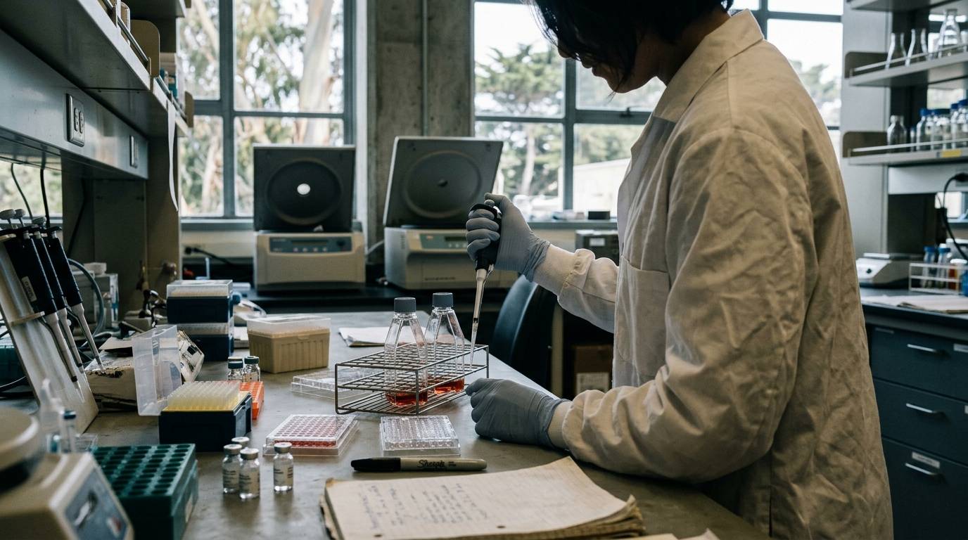
Researchers often search for why a cloned peptide sequence fails to display biological activity when expressed in standard cellular assays. The core problem is that mature peptide hormones and neuropeptides are rarely direct products of simple protein translation. In native cellular biology, active signaling peptides originate as extended, biologically inert precursor proteins. These precursors require ordered compartmental transit, precise enzymatic cleavages, and tailored post-translational modifications before functional signaling molecules emerge.
This guide provides a definitive architectural overview of the eukaryotic secretory pathway responsible for endogenous peptide generation. It maps the pathway from initial ribosomal translation and signal sequence excision to terminal exocytosis and extracellular diffusion. By examining documented biological mechanisms across endocrine and neuroendocrine systems, laboratory researchers can better evaluate precursor processing, identify analytical artifacts, and distinguish mature peptides from intermediate processing fragments.
Precursor Architecture and the Logic of Peptide Biosynthesis
Peptide signals operate across vast physiological distances, coordinating metabolism, vascular tone, neurotransmission, and systemic homeostasis. Despite their structural diversity, most endogenous bioactive peptides share a common biosynthetic origin as prepropeptides or preprohormones. A prepropeptide is an extended precursor polypeptide, commonly containing between 100 and 350 amino acid residues. This extended precursor contains specific architectural domains arranged in a strictly ordered sequence. At the amino terminus sits a hydrophobic signal peptide, which is followed by a prodomain and one or more mature peptide sequences flanked by specialized cleavage motifs.
The evolutionary persistence of precursor processing serves several biological imperatives. Small peptides ranging from three to forty amino acids lack the tertiary structural elements required to establish stable folding states. Without an extended protein scaffold, nascent short chains cannot reliably avoid rapid cytosolic proteolysis. Synthesizing a peptide within a larger precursor polypeptide permits stable thermodynamic folding within the endoplasmic reticulum. This precursor scaffold protects critical structural loops, positions internal disulfide bonds, and sequesters potent bioactive domains until the molecule reaches an appropriate storage organelle.
Furthermore, precursor design enables coordinated stoichiometric production of multiple distinct signaling factors from a single genetic transcript. In polyprotein precursors, a single open reading frame encodes multiple tandem copies of an active peptide or several structurally distinct regulatory hormones. Enzymatic processing can liberate these components simultaneously or selectively, depending on the tissue-specific proteases present within the cell. Consequently, measuring genomic transcription or precursor translation alone does not establish which mature, biologically active molecules a specific cell type actually secretes.
Understanding this distinction is vital for accurate study interpretation. When reviewing published literature, researchers must differentiate between the presence of a propeptide gene and the verified secretion of an active signaling product. Analytical guides on interpreting peptide research literature emphasize that precursor mRNA expression does not automatically guarantee equivalent downstream biological activity. A rigorous evaluation requires confirming the presence of competent processing enzymes alongside the intact mature sequence.
Translation, Endoplasmic Reticulum Entry, and Nascent Chain Folding
The peptide biosynthetic sequence begins in the cytosol, where transfer RNAs deliver amino acids to actively translating ribosomes. As the initial amino-terminal sequence emerges from the ribosomal exit tunnel, it presents a specialized signal peptide. This signal sequence typically comprises 15 to 30 amino acids characterized by a central hydrophobic core flanked by polar residues. The hydrophobic core is recognized by the signal recognition particle, a ribonucleoprotein complex that arrests translation temporarily. The signal recognition particle guides the ribosome-nascent chain complex directly to the membrane of the rough endoplasmic reticulum.
At the endoplasmic reticulum membrane, the complex docks with the signal recognition particle receptor, transferring the nascent polypeptide to the Sec61 translocon pore. Translation then resumes, threading the growing polypeptide chain directly across the lipid bilayer into the endoplasmic reticulum lumen. As the chain enters this luminal environment, a membrane-bound signal peptidase complex cleaves the signal peptide at a designated consensus site. This endoproteolytic excision transforms the preprohormone into a prohormone, releasing the chain to begin its luminal folding trajectory.
Inside the endoplasmic reticulum, the propeptide encounters specialized molecular chaperones and oxidoreductases. Enzymes such as protein disulfide isomerase catalyze the formation and isomerization of covalent disulfide bridges between conserved cysteine residues. For many hormones, correct disulfide formation within the endoplasmic reticulum is an obligatory prerequisite for exit to downstream compartments. The luminal quality control machinery inspects the propeptide for misfolded regions, hydrophobic surface exposure, and inappropriate aggregation.
Propeptides that achieve their native conformational state are packaged into coat protein complex II vesicles. These vesicles bud from specialized endoplasmic reticulum exit sites, ferrying their folded cargo toward the cis-Golgi network. Propeptides that fail these conformational checkpoints are retained by luminal chaperones like calreticulin and BiP. Such misfolded precursors are ultimately retrotranslocated into the cytosol for ubiquitination and proteasomal destruction. The endoplasmic reticulum acts strictly as an entry, folding, and quality-assurance compartment. It is not the site where mature peptide hormones acquire their final endoproteolytic cleavages.
Golgi Compartmentalization and Secretory Pathway Sorting
Upon fusing with the cis-cisternae of the Golgi apparatus, propeptides progress sequentially through the medial and trans-Golgi stacks via vesicular transport or cisternal maturation. As cargo traverses the Golgi complex, it undergoes an array of early post-translational modifications. These events can include core oligosaccharide trimming, complex N-linked or O-linked glycosylation, tyrosine sulfation, and selective serine or threonine phosphorylation. These chemical alterations help protect the protein backbone from premature degradation and modulate its interactions with luminal sorting receptors.
The trans-Golgi network represents the primary organizational junction within the secretory pathway. Here, fully folded and partially modified propeptides are sorted into distinct physical routes based on structural sorting motifs and biophysical properties. The cell maintains two fundamental secretory mechanisms: the constitutive secretory pathway and the regulated secretory pathway. Understanding the biophysical segregation between these two pathways is essential for evaluating peptide production in both native tissues and recombinant host systems.
The constitutive pathway operates continuously in all eukaryotic cells, providing an uninterrupted flow of lipids and proteins directly to the plasma membrane. Vesicles budding via the constitutive pathway do not store cargo for prolonged periods. Instead, they traffic directly to the cell surface, fusing with the membrane without requiring an external physiological trigger. Proteins lacking specific retention or dense-core sorting signals default to this continuous release route.
In contrast, specialized neuroendocrine and endocrine cells direct peptide precursors into the regulated secretory pathway. This specialized route packages propeptides alongside dedicated processing enzymes into nascent secretory organelles. The sorting process at the trans-Golgi network involves cargo condensation, selective receptor interactions, and lipid microdomain segregation. While specific sorting receptors such as carboxypeptidase E have been implicated for certain cargoes, extensive research suggests that homotypic cargo aggregation driven by luminal environments represents the principal sorting driver. For deeper background on cell-specific mechanisms, researchers can review curated materials covering peptide fundamentals and biology.
Dense-Core Granule Biogenesis and Acidification Dynamics
The defining organelle of the regulated secretory pathway is the dense-core granule, also termed the large dense-core vesicle in neurobiology. Dense-core granules originate as immature secretory granules budding from the trans-Golgi network. Electron microscopy reveals these immature organelles as electron-dense protein cores enclosed within a loosely fitting lipid bilayer membrane. Biogenesis of these structures depends heavily on granin family proteins, including chromogranin A, chromogranin B, and secretogranin II. Granins possess an intrinsic capacity to condense into insoluble macromolecular aggregates under specific luminal conditions.
As immature secretory granules pinch off from the trans-Golgi network, they undergo an extensive maturation process. Maturation requires three coordinated physical events: progressive luminal acidification, selective membrane remodeling, and the spatial condensation of core cargo. Newly formed immature granules maintain a near-neutral internal pH of roughly 6.5. As they migrate along the cytoskeleton, vacuolar-type proton adenosine triphosphatases embedded within the granule membrane actively pump cytosolic protons into the granule lumen. This proton accumulation steadily drives the internal pH down toward an optimal range of 5.0 to 5.5.
Concurrently, the immature granule expels membrane proteins and fluid that are not intended for long-term storage. Clathrin-coated patches bud from the immature granule membrane, recycling excess membrane, syntaxins, and mis-sorted constitutive proteins back to the trans-Golgi network or endosomal compartments. This loss of surface area and fluid forces the internal protein cargo into an ultra-dense, semi-crystalline matrix.
This tight physical packaging serves two critical biological functions. First, it permits cells to store peptide concentrations reaching tens of millimolar within a restricted intracellular volume. Second, the progressive reduction in pH, accompanied by elevated luminal calcium concentrations, directly activates dormant prohormone convertases co-packaged within the granule. Thus, granule maturation coordinates organelle remodeling with the temporal activation of downstream processing enzymes.
Endoproteolytic Processing and the Prohormone Convertase Cascade
The conversion of an inert prohormone into an active signaling peptide requires site-specific endoproteolytic cleavage. The primary enzymes executing these cleavages belong to the proprotein convertase subtilisin/kexin family. Within regulated neuroendocrine granules, two calcium-dependent serine endoproteases predominate: prohormone convertase 1/3 and prohormone convertase 2. Both enzymes share evolutionary homology with bacterial subtilisin and yeast kexin. However, they exhibit distinct pH optima, calcium dependencies, and cleavage site preferences.
Prohormone convertase 1/3 and prohormone convertase 2 recognize specific motifs containing basic amino acid residues. Classical cleavage sites typically comprise pairs of basic residues, such as arginine-arginine, lysine-arginine, arginine-lysine, or lysine-lysine. Cleavage occurs immediately at the carboxyl-terminal side of the basic pair. In certain structural contexts, prohormone convertases can also recognize monobasic sites or consensus motifs defined by basic residues separated by several spacer amino acids.
The activation of these convertases is strictly regulated in time and space to prevent premature cleavage within early secretory compartments. Prohormone convertase 1/3 is synthesized as an inactive zymogen that undergoes an initial autocatalytic cleavage within the neutral endoplasmic reticulum. However, its prodomain remains non-covalently bound, acting as an autoinhibitor until the enzyme reaches the acidic, calcium-rich environment of the maturing secretory granule. At pH 5.5, a secondary internal cleavage destroys the prodomain, releasing the fully active enzyme.
Prohormone convertase 2 follows an even more intricate maturation pathway. Its initial autoactivation requires binding to a specialized neuroendocrine chaperone known as 7B2. The 7B2 protein prevents premature aggregation of prohormone convertase 2 within the endoplasmic reticulum. It facilitates transit to the trans-Golgi network, where cleavage of 7B2 permits prohormone convertase 2 to autoactivate under acidic granule conditions.
Because prohormone convertase 1/3 activates at a slightly higher pH than prohormone convertase 2, processing inside the maturing granule often follows an ordered chronological sequence. Prohormone convertase 1/3 typically initiates the first endoproteolytic cut on the precursor. As the granule acidifies further, prohormone convertase 2 executes secondary cleavages on the resulting intermediate fragments. This sequential proteolysis ensures that complex precursors are processed in a reproducible, stepwise manner.
Post-Translational Trimming, Terminal Amidation, and Structural Stabilization
Endoproteolytic cleavage by prohormone convertases does not yield a finished signaling molecule. When a convertase cleaves at a dibasic recognition site, the resulting amino-terminal peptide fragment retains basic arginine or lysine residues attached to its carboxyl terminus. These positively charged residues disrupt receptor-binding interactions and destabilize the molecule. To resolve this, cells deploy exopeptidases to polish the terminal ends.
Carboxypeptidase E is the primary metallo-exopeptidase responsible for trimming these residual basic amino acids. Operating optimally within the acidic granule environment, carboxypeptidase E sequentially removes terminal lysine and arginine residues one by one. The enzyme halts its exopeptidase action as soon as it exposes a non-basic residue. The resulting peptide features its native carboxyl-terminal sequence, but in many cases, it still requires terminal chemical modification to achieve biological potency.
A major modification found across neuropeptides and endocrine signals is carboxyl-terminal amidation. In unamidated peptides, the terminal carboxyl group carries a negative charge under physiological pH. Amidation replaces this terminal hydroxyl group with an uncharged amide group. This modification neutralizes the carboxyl-terminal charge, mimicking a continuing peptide backbone. Amidation protects the peptide from systemic exopeptidase degradation and is frequently required for high-affinity docking with target G-protein coupled receptors.
Carboxyl-terminal amidation is performed by peptidylglycine alpha-amidating monooxygenase, a bifunctional enzyme anchored in the granule membrane or present in soluble form. This complex enzyme executes a two-step catalytic reaction:
- Peptidyl alpha-hydroxylating monoxygenase consumes molecular oxygen, copper ions, and ascorbic acid to hydroxylate the alpha-carbon of an obligatory carboxyl-terminal glycine residue.
- Peptidylamidoglycolate lyase catalyzes the zinc-dependent cleavage of the intermediate, releasing glyoxylate and leaving behind an amidated carboxyl terminus.
Beyond amidation, peptides can undergo amino-terminal modifications that shield them from aminopeptidase digestion. An example is the conversion of an exposed amino-terminal glutamine or glutamate residue into pyroglutamate, catalyzed by glutaminyl cyclase. Other peptides require specific amino-terminal acetylation, tyrosine sulfation, or aliphatic acylation to enable receptor activation. Each modification demands precise spatial coordination with endoproteolytic processing.
Laboratory verification of synthetic or isolated research peptides must account for these complex terminal chemistry details. When evaluating research peptide identity, investigators should confirm the exact terminal state using high-resolution analytical documentation. The role of orthogonal mass spectrometry and chromatographic profiling is detailed in laboratory guides on reading peptide certificates of analysis.
Comparative Biosynthetic Models: Insulin, Proglucagon, and Proopiomelanocortin
Examining how native tissues handle specific propeptides illustrates the cellular logic of precursor processing. Cells do not follow a uniform, monolithic script. Instead, they leverage precursor sequence architecture and differential enzyme expression to generate tissue-specific peptide arrays.
- Preprohormone Architecture (Generic)
- Signal Peptide
- N-terminal Prodomain
- Cleavage Site
- Mature Peptide
- C-terminal Prodomain
- Proinsulin Processing Cascade
- Preproinsulin (110 aa) (Signal Peptidase, ER) Proinsulin (B-chain - C-peptide - A-chain)
- Disulfide Bonding (ER Lumen)
- Packaging into Dense-Core Granule
- PC1/3 Cleaves at B-chain / C-peptide junction PC2 Cleaves at C-peptide / A-chain junction
- Carboxypeptidase E Trimming
- (Removes Arg-Arg / Lys-Arg)
- Mature Insulin (A-B Heterodimer) Free C-Peptide
Proinsulin and Two-Chain Heterodimer Assembly
Insulin provides the classic model for endoproteolytic maturation within pancreatic beta cells. Preproinsulin is translated as a 110-amino acid single-chain precursor. Its amino-terminal signal peptide guides it into the endoplasmic reticulum, where signal peptidase cleaves it to form proinsulin. Proinsulin consists of an amino-terminal B-chain, an internal connecting peptide known as C-peptide, and a carboxyl-terminal A-chain.
Within the endoplasmic reticulum, proinsulin folds so that the B-chain and A-chain align, stabilized by the C-peptide domain. Protein disulfide isomerase catalyzes the formation of two interchain disulfide bonds between the A and B chains, alongside an intrachain disulfide bond within the A-chain. Proinsulin is subsequently sorted into immature secretory granules.
Within the maturing beta-cell granule, prohormone convertase 1/3 and prohormone convertase 2 work cooperatively. Prohormone convertase 1/3 initiates cleavage at the junction between the B-chain and the C-peptide. Carboxypeptidase E then removes the exposed basic residues. Subsequently, prohormone convertase 2 cleaves the junction between the C-peptide and the A-chain, and carboxypeptidase E removes the remaining basic residues.
This process yields mature insulin, a 51-amino acid heterodimer linked by disulfide bonds, along with an equimolar quantity of free C-peptide. Measuring mature insulin without monitoring proinsulin processing intermediates can produce misleading analytical findings in beta-cell biology. Intermediate fragments possess vastly reduced metabolic activity yet may cross-react with poorly validated immunoassays.
Proglucagon and Tissue-Specific Cleavage Divergence
Proglucagon demonstrates how identical precursor transcripts produce distinct biological signals depending on the host cell type. The proglucagon gene is expressed in both pancreatic alpha cells and neuroendocrine L cells of the distal intestine. The translated proglucagon precursor contains the amino acid sequences for glucagon, glucagon-like peptide 1, glucagon-like peptide 2, and several intervening peptide domains.
In pancreatic alpha cells, the processing machinery expresses high levels of prohormone convertase 2 and minimal prohormone convertase 1/3. Under these conditions, prohormone convertase 2 cleaves the precursor to liberate:
- Mature glucagon, a 29-amino acid hormone that stimulates hepatic glycogenolysis.
- Glicentin-related pancreatic polypeptide.
- A large, unprocessed carboxyl-terminal fragment known as the major proglucagon fragment.
In intestinal L cells, the cellular processing complement is reversed. L cells express robust quantities of prohormone convertase 1/3 while lacking prohormone convertase 2. Prohormone convertase 1/3 ignores the internal cleavage sites required to liberate glucagon. Instead, it cleaves proglucagon to generate:
- Glucagon-like peptide 1, which potentiates glucose-dependent insulin secretion.
- Glucagon-like peptide 2, which promotes mucosal epithelial proliferation.
- Oxyntomodulin and glicentin.
Thus, finding proglucagon mRNA within a tissue or cell line does not clarify what signal is actually being produced. The identity of the secreted product is dictated entirely by which convertase enzyme the cell expresses.
Proopiomelanocortin and Multifunctional Polyprotein Cleavage
Proopiomelanocortin represents the archetype of a multifunctional polyprotein precursor. Expressed primarily in the corticotrope cells of the anterior pituitary and proopiomelanocortin neurons of the arcuate nucleus, this single precursor contains sequences for multiple distinct signaling molecules.
In the anterior pituitary, corticotropes express prohormone convertase 1/3. This enzyme cleaves proopiomelanocortin into adrenocorticotropic hormone, an essential regulator of adrenal corticosteroid synthesis, and beta-lipotropin. The cell does not execute further endoproteolytic cleavage on adrenocorticotropic hormone.
In the intermediate pituitary lobe and hypothalamic neurons, cells express both prohormone convertase 1/3 and prohormone convertase 2. In these tissues, prohormone convertase 2 cleaves adrenocorticotropic hormone further, producing alpha-melanocyte-stimulating hormone and corticotropin-like intermediate peptide. Subsequent enzymes amidate alpha-melanocyte-stimulating hormone and acetylate its amino terminus. Simultaneously, beta-lipotropin is cleaved to yield beta-endorphin, an endogenous opioid peptide. Proopiomelanocortin processing illustrates how cellular enzymatic profiles can completely alter the physiological class of the resulting signaling output.
Stimulus-Coupled Exocytosis and Non-Synaptic Peptide Diffusion
Once dense-core granules complete their maturation, they do not spontaneously fuse with the plasma membrane. Instead, they are transported along microtubule tracks via kinesin motor proteins to the cell periphery. At the actin-rich cell cortex, granules transition to myosin-driven transport and enter a regulated exocytotic cycle. This cycle is physically orchestrated by soluble N-ethylmaleimide-sensitive factor attachment protein receptor complexes, commonly termed SNARE proteins.
The granule membrane incorporates vesicular SNARE proteins, primarily synaptobrevin-2 or cellubrevin. The plasma membrane contains target SNARE proteins, comprising syntaxin-1 and synaptosome-associated protein 25. As granules approach the cell boundary, these complementary SNARE proteins intertwine into a tight four-helix bundle. This assembly pulls the granule membrane into close proximity with the plasma membrane, a state known as vesicle docking and priming.
Exocytosis is triggered by a rise in free intracellular calcium. In endocrine cells and neurons, membrane depolarization or receptor-mediated signaling opens voltage-gated calcium channels or stimulates inositol trisphosphate-mediated release from intracellular stores. The calcium sensor synaptotagmin, embedded in the granule membrane, binds these incoming calcium ions. This binding induces synaptotagmin to interact with the SNARE complex and acidic phospholipids, driving immediate fusion pore opening and contents release.
Peptide release dynamics differ substantially from classical small-molecule neurotransmission:
- Dense-core granules often require prolonged, high-frequency stimulation or global intracellular calcium elevations to trigger exocytosis, whereas small synaptic vesicles release cargo following localized, transient calcium microdomains.
- Neuropeptide release is not restricted to active synaptic zones. Dense-core granules can undergo exocytosis from neuronal soma, dendrites, and non-synaptic axonal varicosities.
- Peptides are stored as concentrated matrices that dissolve more slowly upon pore opening, producing sustained release profiles rather than instantaneous quantal bursts.
Following non-synaptic exocytosis, neuropeptides diffuse through the interstitial fluid to engage high-affinity G-protein coupled receptors on adjacent or distant cells. This diffuse communication mode, known as volume transmission, allows peptides to modulate neural circuits across large tissue volumes.
Signal termination also diverges fundamentally from classical neurotransmitter systems. Cells do not possess high-affinity reuptake transporters for released peptides. Instead, signaling ceases via passive extracellular diffusion and enzymatic proteolysis by cell-surface metalloendopeptidases, such as neprilysin and insulin-degrading enzyme. In some physiological contexts, these extracellular cleavage events do not simply destroy the signal. Instead, they clip the peptide into shorter secondary fragments that display altered receptor selectivity.
Critical Unknowns, Methodological Limitations, and Experimental Nuances
Despite extensive characterization of the general secretory pathway, significant mechanistic questions and analytical challenges remain in peptide biology. Researchers studying endogenous peptides or working with cell culture models must navigate several confounding factors.
First, sorting mechanisms at the trans-Golgi network remain an active area of investigation. While cargo aggregation driven by low pH and high calcium is widely observed, the precise balance between receptor-mediated sorting and passive physical condensation varies considerably between cell types. Investigators cannot assume that expressing a human propeptide within a non-endocrine mammalian cell line will direct the cargo into a regulated secretory pathway. In cells lacking dense-core machinery, precursors are routinely diverted into the constitutive pathway, leading to premature secretion of unprocessed, inactive proforms.
Second, estimating the physical peptide content per dense-core granule involves notable experimental uncertainty. Published reviews estimate peptide concentrations within mature granules at approximately 3 to 10 millimolar. This equates to roughly 10,000 peptide molecules per vesicle in model neuroendocrine systems. However, these estimates rely on idealized mathematical assumptions regarding spherical volume and matrix packaging. Extrapolating quantitative parameters from densely granulated hypothalamic magnocellular neurons to peripheral endocrine cells or primary cell lines can introduce significant calculation errors.
Third, extrapolating systemic pharmacokinetic half-life to local tissue signaling persistence represents a frequent interpretive pitfall. Researchers often cite a peptide's plasma half-life as an absolute metric of its biological duration. However, plasma clearance values reflect systemic hepatic filtration, renal clearance, and intravascular peptidase activity. They do not accurately describe the longevity of a peptide signal within the brain parenchyma, cerebrospinal fluid, or dense interstitial matrix. In localized tissue compartments, shielding from soluble peptidases or slow dissolution from extracellular matrices can extend biological signaling far beyond systemic elimination timelines.
Finally, researchers must recognize the translational divide separating preclinical cellular models from human physiology. Cell-free assays and immortalized cell lines frequently express aberrant ratios of prohormone convertases compared to primary human tissues. A complete scientific evaluation requires inspecting multiple levels of empirical evidence. A structured overview of these analytical tiers can be found in our framework on peptide evidence levels and study quality. Researchers should never assume that an endoproteolytic cleavage observed in an engineered cell model reproduces the processing dynamics of intact native tissue.
Analytical Verification Checklist for Laboratory Researchers
When designing laboratory experiments, evaluating research papers, or sourcing peptide reference standards, researchers should apply a systematic checklist to verify compound identity and biological relevance. Use this seven-step verification sequence to inspect published claims and experimental designs:
- Identify the precursor status of your target signal. Determine whether the experimental system evaluates the full preprohormone, the propeptide intermediate, or the fully processed mature peptide chain.
- Characterize the host cell processing machinery. Confirm that the model cell line expresses the necessary prohormone convertases, such as prohormone convertase 1/3 or 2, alongside required auxiliary proteins like 7B2.
- Verify terminal post-translational modifications. Check whether the target sequence requires carboxyl-terminal amidation, amino-terminal pyroglutamylation, or internal disulfide bonds for receptor binding.
- Distinguish regulated storage from constitutive leakage. Measure whether peptide secretion is coupled to calcium-dependent secretagogues, or whether baseline medium accumulation indicates constitutive default secretion.
- Inspect the spatial site of release. When working with neuronal models, determine whether peptide release occurs across synaptic junctions, axonal varicosities, or dendritic compartments.
- Verify reference standard specifications. When acquiring research-use peptides, review the high-performance liquid chromatography and mass spectrometry data to ensure the synthesized sequence matches the processed mature hormone rather than a truncated intermediate. For sequence terminology conventions, consult our guide on peptide names, sequences, and compound identity.
- Check antibody and assay cross-reactivity. Confirm whether the analytical detection methods employed, such as enzyme-linked immunosorbent assays or radioimmunoassays, cross-react with unprocessed proforms or degradation fragments.
By approaching peptide biosynthesis through this structured cellular framework, researchers can accurately differentiate mature bioactive signals from immature precursor intermediates, preventing costly interpretive errors in laboratory peptide science.
Sources
- Overview of neuropeptides: awakening the senses? - PMC - NIH
- Biochemistry, Peptide - StatPearls - NCBI Bookshelf - NIH
- Heterogeneous Expression of Proinsulin Processing Enzymes ...
- Biochemistry, Hormones - StatPearls - NCBI Bookshelf - NIH
- Severe block in processing of proinsulin to insulin ...
- The making of insulin in health and disease | Diabetologia
- Understanding peptide hormones: from precursor proteins to bioactive molecules
- The Insulin Monomer
- Neuropeptide Transmitters: Biosynthesis Release and Response - Studocu
Introducing / CaliLean Field Notes
A little more
perspective.
An editorial letter for curious minds. Thoughtful reading, questions worth keeping and the sources behind them.
Explore Field Notes Join the list for the first edition

