
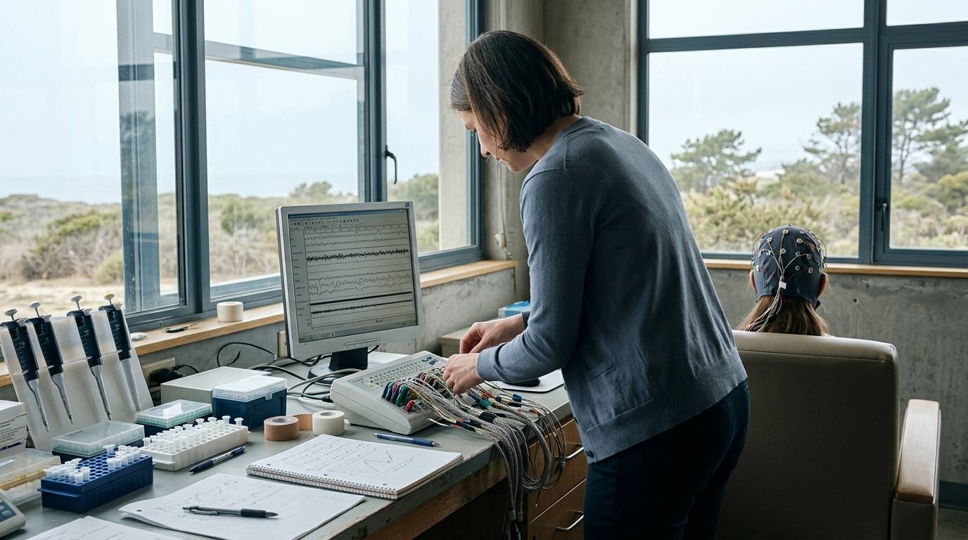
Many laboratory protocols treat physical movement as a direct proxy for wakefulness. This assumption is fundamentally flawed. An animal can engage in stereotyped running while exhibiting slow-wave cortical dynamics. Conversely, an animal can remain completely stationary while experiencing heightened cortical vigilance and autonomic activation.
In preclinical peptide research, conflating motor output with vigilance leads to misclassified phenotypes and irreproducible findings. Investigating candidate neuropeptides requires distinguishing between behavioral movement, global sleep-wake states, and fine-grained neural arousal.
Rigorous research demands validated electrophysiological recordings paired with multi-parametric behavioral and physiological monitoring. Researchers studying candidate molecules in brain, sleep, and cognitive peptide research must isolate genuine shifts in vigilance from motor confounds, procedural stress, and circadian baselines.
Distinguish Brain State, Arousal Intensity, and Motor Activity
Investigating arousal pathways begins by separating three distinct biological phenomena: sleep-wake state, arousal intensity, and motor activity. Wakefulness is an electrophysiologically scored brain-body state classified through continuous electroencephalography (EEG) and electromyography (EMG). Arousal represents a broader physiological construct that spans cortical desynchronization, autonomic tone, sensory responsiveness, and heightened attention. Locomotion is simply an overt motor behavior that frequently accompanies active waking.
A candidate peptide might stimulate spontaneous motor activity without altering baseline sleep architecture. Another molecule might prolong cortical desynchronization while the animal remains motionless in a resting posture. When researchers rely solely on infrared beam breaks or video-tracking software, these divergent neurobiological profiles appear identical.
To prevent misinterpretation, researchers structure their experimental protocols across four distinct operational levels. The first level identifies the categorical macro-state: wake, non-rapid eye movement (NREM) sleep, or rapid eye movement (REM) sleep. The second level evaluates state quality by analyzing spectral power distributions, bout durations, and micro-architectural fragmentation.
The third level records circuit-specific activity within defined neuroanatomical pathways, such as the lateral hypothalamus or locus coeruleus. The fourth level evaluates behavioral and peripheral physiology, including pupillometry, posture, and autonomic tone. Examining each layer independently allows scientists to determine whether a peptide alters vigilance systems directly or merely modifies secondary behavioral outputs.
Implement Polysomnography to Establish Objective Sleep-Wake Stages
Polysomnography remains the gold standard for defining sleep-wake architecture in mammalian models. In rodent studies, researchers implant chronic cranial electrodes to record cortical EEG alongside bilateral nuchal electrodes to monitor skeletal muscle EMG. Signals must be sampled at rates exceeding 100 Hz to prevent aliasing and capture high-frequency components.
The recorded data is partitioned into short, discrete epochs, typically lasting between two and ten seconds. Each epoch is assigned to a discrete state based on validated electrophysiological characteristics. Wakefulness displays low-amplitude, high-frequency EEG patterns paired with variable, tonically elevated EMG activity.
NREM sleep, often termed slow-wave sleep in animal models, displays high-amplitude slow-wave activity dominated by delta rhythms between 0.5 and 4.0 Hz, accompanied by reduced muscle tone. REM sleep presents a distinct signature marked by rhythmic, high-frequency theta activity between 6.0 and 9.0 Hz alongside profound skeletal muscle atonia. Without simultaneous EMG recording, theta-dominated REM sleep can be easily misclassified as active wakefulness.
Modern laboratories frequently combine automated scoring algorithms with blinded manual review. Automated classifiers typically apply an empirical EMG threshold to separate sleep from wakefulness, followed by an EEG theta-to-delta power ratio to differentiate NREM from REM sleep. Because arbitrary threshold selections alter cumulative state totals, researchers must explicitly report their calibration rules and scoring parameters. Adhering to strict evidence levels in peptide research requires full transparency regarding how raw bio-signals are filtered, binned, and verified.
Differentiate Active Wake from Quiet Wake in Experimental Designs
The waking state is not a homogeneous physiological condition. Preclinical investigations must separate active wakefulness from quiet wakefulness. Active wake involves gross motor locomotion, exploratory rearing, grooming, and high muscle tone. Quiet wake involves awake immobility, characterized by resting posture, absent locomotion, reduced EMG amplitude, and preserved sensory responsiveness.
This biological distinction is particularly evident in peptidergic circuits that govern vigilance. Lateral hypothalamic neurons that synthesize orexin (also known as hypocretin) fire vigorously during active waking when muscular exertion and exploratory locomotion occur. During quiet waking, the discharge rate of these identical peptidergic neurons decreases dramatically, falling to near-silent levels during NREM and REM sleep.
Their discharge correlates positively with fast gamma EEG rhythms and nuchal EMG activity during wakefulness. Conversely, their firing correlates negatively with slow delta oscillations. If an assay fails to distinguish quiet wake from sleep, it will systematically underestimate total wake time.
Similarly, if an assay counts only locomotor bouts, it will mischaracterize quiet wake as inactive or sedated states. For instance, microinjections of orexin-A into the rostral lateral hypothalamus of rodent models elicit significant increases in active waking alongside reductions in slow-wave and REM sleep. Yet the duration of quiet waking during the initial post-injection hour remains entirely unchanged. Studies that aggregate active and quiet wake into a single metric miss these critical behavioral patterns.
Quantify Sleep Fragmentation and Define Transient Micro-Arousals
Evaluating a peptide requires examining more than total cumulative minutes spent awake. Researchers must quantify sleep architecture, state continuity, and transient micro-arousals. A drug or peptide might leave total 24-hour wake time unchanged while severely destabilizing state maintenance, causing profound fragmentation.
In clinical human sleep scoring, an arousal requires an abrupt frequency shift in the EEG montage lasting at least three seconds. This event must be preceded by at least ten continuous seconds of stable sleep. When scoring human REM sleep, this EEG acceleration must also be accompanied by a concurrent increase in submental EMG tone lasting at least one second.
Translational rodent experiments cannot apply human scoring rules without modification. Rodent sleep-wake cycles are polyphasic and organized into much shorter bouts. However, rodent researchers must still operationalize their definitions of micro-arousal, awakening, and state transitions with rigorous precision.
Researchers assess micro-architectural stability by extracting several key parameters:
- Sleep latency: the duration from compound administration to the onset of the first consolidated sleep bout.
- Mean bout duration: the average continuous length of individual wake, NREM, and REM episodes.
- State transition frequency: the total number of shifts between distinct vigilance states per recording window.
- Brief arousal frequency: transient EEG frequency accelerations lasting between one and five seconds that do not progress into consolidated wake bouts.
Movement alone does not constitute an electrophysiological arousal event. Peripheral muscle twitches and brief myoclonic jerks occur routinely during healthy rodent REM and NREM sleep without disrupting underlying cortical slow waves. Equating automated piezoelectric or accelerometer movements with cortical awakening introduces significant scoring bias.
Map Neural Circuit Dynamics Alongside Electrophysiological Recording
To understand how neuropeptides regulate vigilance, researchers map specific neural circuits using modern genetic and physiological tools. Investigating pathways such as orexinergic, histaminergic, or noradrenergic systems requires observing how defined cell types respond during spontaneous and stimulated states. Optogenetic, chemogenetic, and fiber photometric approaches permit cell-type-specific interrogation in vivo.
Optogenetic stimulation experiments show that selective photostimulation of orexin-expressing neurons in the lateral hypothalamus increases the probability of transitioning from sleep to wakefulness. Conversely, optogenetic inhibition of this cell group suppresses wakefulness and increases NREM sleep time. These circuit manipulations demonstrate functional sufficiency and necessity within endogenous arousal networks.
However, manipulating neural circuits yields interpretable data only when paired directly with simultaneous polysomnography. Fiber optic cables and gradient-index lenses must be chronically integrated alongside cranial EEG screws and nuchal EMG wires. Measuring calcium transients or multi-unit firing in isolation reveals that a circuit is active, but it cannot confirm that the animal has transitioned into physiological wakefulness.
Targeted pharmacology demonstrates similar pathway interactions. For example, the wake-promoting efficacy of centrally administered orexin-A is attenuated in mice lacking functional histamine H1 receptors or in wild-type animals pretreated with H1-receptor antagonists. This demonstrates that downstream histaminergic signaling from the tuberomammillary nucleus is critical for mediating peptide-driven arousal. Researchers must avoid substituting single-cell firing rates or regional calcium fluorescence for validated state scoring.
Incorporate Pupil Dynamics and Autonomic Signals Without Conflating Measures
To capture changes in arousal intensity beyond categorical sleep stages, investigators frequently track pupillometry and autonomic parameters. Pupil diameter serves as a sensitive, noninvasive real-time readout of central noradrenergic and cholinergic tone. Fluctuations in pupil size track activity within the locus coeruleus and basal forebrain, providing a high-resolution window into internal brain state.
Pupil diameter correlates reliably with locomotion and active exploration. When an animal runs or actively sniffs, its pupils dilate substantially. However, pupil diameter also exhibits meaningful fluctuations while animals remain completely stationary. These baseline variations reveal internal shifts in central arousal and sensory processing that cannot be detected by video tracking alone.
Autonomic metrics provide further insight into physiological activation. Continuous recording of heart rate, arterial blood pressure, and core body temperature via implantable telemetry reveals changes in sympathetic and parasympathetic balance. Pharmacological agents and peptides can trigger prominent cardiovascular activation, elevating heart rate and blood pressure during sleep periods without inducing overt cortical awakenings.
These autonomic markers must be treated as complementary readouts rather than standalone measures of wakefulness. An increase in heart rate or transient pupil dilation reflects autonomic arousal, but it does not prove that an animal has achieved EEG-defined wakefulness. When interpreting preclinical peptide literature, readers must verify that investigators kept autonomic tone, pupil metrics, and cortical sleep staging methodologically distinct.
Control for Injection Stress, Circadian Timing, and Experimental Confounders
A common flaw in preclinical peptide research is the failure to control for external experimental variables that mimic or obscure physiological wakefulness. The physical act of handling an animal, applying physical restraint, and administering an injection elicits acute stress. Stress activates the sympathetic nervous system and the hypothalamic-pituitary-adrenal axis, releasing endogenous corticotropin-releasing factor and catecholamines that trigger immediate behavioral arousal.
If an investigator injects a candidate peptide and observes two hours of continuous wakefulness, that wakefulness may stem entirely from injection stress rather than peptide pharmacology. To eliminate this confound, protocols must incorporate extensive animal habituation to handling and recording tethers. Vehicle-controlled crossover designs are essential, where each animal receives vehicle and active compound at identical circadian points under identical procedural conditions.
Circadian timing represents another vital experimental variable that dictates baseline sleep pressure. Mammals exhibit distinct biological rhythms governed by the suprachiasmatic nucleus. In nocturnal rodents, the light phase is characterized by high sleep propensity, whereas the dark phase is characterized by consolidated wakefulness.
A wake-promoting peptide administered during the early light phase (for example, Zeitgeber Time 3) acts against high homeostatic sleep pressure, making its wake-promoting properties readily detectable. If the same compound is administered at dark-phase onset, when baseline wakefulness is already elevated near its ceiling, its wake-promoting properties may appear negligible. Researchers must fully report zeitgeber timing, photoperiod conditions, and baseline sleep architecture.
Methodological reporting should adhere strictly to the ARRIVE 2.0 guidelines:
- State the exact species, strain, sex, age, and microbiological status of the experimental subjects.
- Report randomization protocols and describe blinding across compound administration, sleep scoring, and spectral data analysis.
- Detail the administration route, precise injection volume, stereotaxic coordinates, and microinjection speed for central deliveries.
- Document animal housing standards, including light-dark cycles, ambient room temperature, and humidity parameters.
Controlling these variables ensures that measured changes in wakefulness reflect the pharmacological properties of the peptide rather than environmental disruptions.
Verify Peptide Sample Identity and Chemical Integrity Before In Vivo Testing
In vivo neurobiological assays rely entirely on the chemical integrity and purity of the test compound. A behavioral or electrophysiological assay cannot yield reliable data if the administered peptide contains truncated sequences, chemical degradation products, or endotoxin contamination. Impurities can introduce biological artifacts that alter neural firing or trigger immune-mediated somnolence.
Synthesizing research-use-only peptides generates crude mixtures containing missing-sequence peptides, diastereomers, and chemical adducts. Investigators must demand rigorous analytical documentation before preparing solutions for central or peripheral administration. High-Performance Liquid Chromatography (HPLC) establishes chromatographic purity, while Mass Spectrometry (MS) verifies that the molecular mass matches the theoretical structure.
Researchers must understand what an analytical report does and does not prove. When evaluating a peptide certificate of analysis, the document verifies only the specific vial or batch tested by the analytical laboratory. It does not ensure long-term stability, correct reconstitution practices, or freedom from post-analytical degradation.
Peptides can degrade rapidly when exposed to improper pH, repeated freeze-thaw cycles, or prolonged storage in aqueous vehicles. Endotoxin testing is especially critical for sleep-wake research, because trace lipopolysaccharide contamination induces robust increases in NREM sleep and disrupts normal slow-wave activity. Maintaining stringent peptide purity and content standards protects behavioral studies from false pharmacological signals.
Avoid Critical Analytical Errors in Preclinical Wakefulness Studies
To design robust sleep-wake experiments, researchers must avoid several pervasive methodological errors that frequently undermine preclinical publications. These errors distort findings, reduce translational utility, and lead to unjustified conclusions regarding peptide efficacy.
Relying Solely on Total Wake Time
Reporting only cumulative wake duration over a multi-hour window conceals important underlying neurobiology. A compound that induces four hours of fragmented, hyper-aroused wakefulness with frequent micro-awakenings produces the exact same total wake time as a compound that supports a single consolidated, calm wake bout. Researchers must report bout length distributions, transition frequencies, and sleep latencies alongside cumulative time-in-state.
Misinterpreting Locomotion as Cortical Desynchronization
Automated open-field boxes and home-cage activity monitors measure physical movement, not brain state. An animal undergoing intense environmental stress may exhibit motor freezing while its brain displays continuous low-voltage, high-frequency waking EEG. Measuring locomotion without polysomnography provides behavioral data, but it cannot support claims regarding sleep-wake regulation.
Ignoring Spectral Power Analysis
Time spent in a state reveals nothing about the electrophysiological quality of that state. Analyzing EEG power spectral density via Fast Fourier Transform provides quantitative insight into cortical dynamics. Examining delta power (0.5 to 4.0 Hz) during NREM sleep tracks homeostatic sleep pressure, while analyzing theta power (6.0 to 9.0 Hz) and gamma power (30 to 80 Hz) during wakefulness reveals the depth of cortical activation. Agonists acting at wake-promoting receptors often alter specific frequency bands without producing proportional shifts in macro-state duration.
Generalizing Preclinical Rodent Findings Across Species
Rodents are polyphasic sleepers that distribute sleep bouts across both the light and dark cycles. In contrast, healthy adult humans are monophasic sleepers whose sleep is consolidated into a single nocturnal block. Furthermore, the metabolic clearance, blood-brain barrier permeability, and receptor distribution of neuropeptides vary considerably across mammalian species. Preclinical findings obtained in rodent models provide valuable mechanistic hypotheses, but they cannot be treated as evidence of human physiological outcomes.
Key Takeaways
- Sleep-wake state, arousal intensity, and locomotor activity represent distinct neurobiological processes that must be measured independently using dedicated tools.
- Continuous polysomnography combining EEG and EMG remains the only validated method for establishing objective wake, NREM, and REM macro-states.
- Waking behavior comprises biologically distinct active and quiet states that exhibit profoundly different peptidergic and electrophysiological profiles.
- Micro-arousals and sleep fragmentation must be explicitly operationalized using validated electrophysiological criteria rather than inferred from transient motor twitches.
- Circuit-level technologies like optogenetics and fiber photometry yield actionable sleep-wake insights only when paired with concurrent polysomnographic recording.
- Noninvasive autonomic markers and pupillometry provide valuable indexes of internal arousal tone, but they cannot substitute for direct cortical state scoring.
- Procedural handling stress, circadian timing, and ambient recording environments represent substantial confounds that require strict vehicle controls and ARRIVE-compliant reporting.
- Peptide integrity, chromatographic purity, and endotoxin verification must be analytically confirmed to prevent chemical and immune artifacts in vivo.
Careful experimental design and rigorous multi-parametric recordings remain the only path to producing reliable, reproducible insights in sleep and arousal research.
Sources
- Wakefulness regulation of memory encoding and retrieval: structure and activity
- Sleep and vigilance states: Embracing spatiotemporal dynamics
- Neuroanatomical Basis for the Orexinergic Modulation of Anesthesia Arousal and Pain Control
- Neural Circuitry of Wakefulness and Sleep - PMC - NIH
- Mechanisms underlying obesity resistance associated with high spontaneous physical activity
- Arousal effect of orexin A depends on activation of the histaminergic system
- Behavioral/Systems/Cognitive
- Scoring Arousals (A) - Sleep ISR
- Disorders of Arousal in adults: new diagnostic tools for ...
- Neurobiology of the Sleep-Wake Cycle: Sleep Architecture, Circadian Regulation, and Regulatory Feedback
- Differential Roles of Orexin Receptor-1 and -2 in the Regulation of Non-REM and REM Sleep
- Arousal and locomotion make distinct contributions to cortical
Introducing / CaliLean Field Notes
A little more
perspective.
An editorial letter for curious minds. Thoughtful reading, questions worth keeping and the sources behind them.
Explore Field Notes Join the list for the first edition

