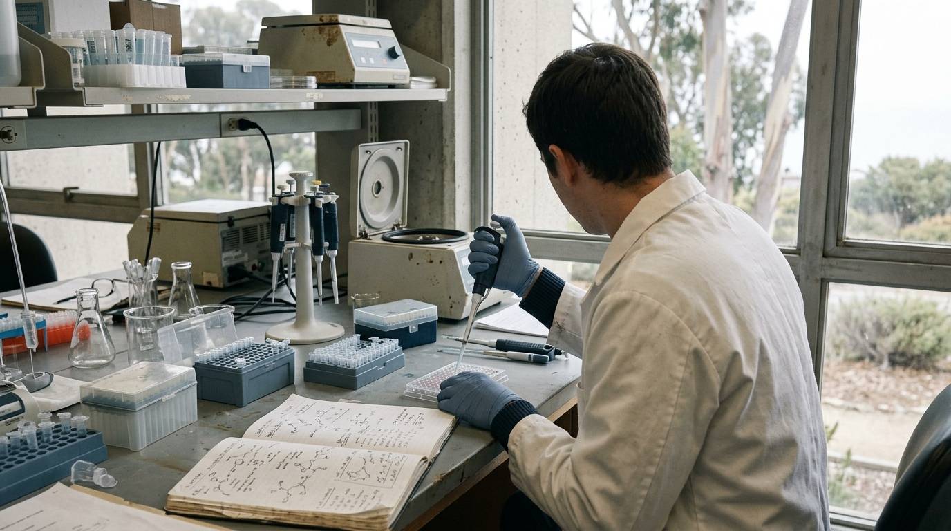
You review a published study that reports a dramatic spike in circulating peptide concentrations. The figures appear definitive at first glance. When you examine the methods section, you notice the chosen assay cross-reacts with inactive metabolic fragments. What looked like a surge in intact, bioactive hormone was partly an accumulation of breakdown products.
Metabolic peptide research requires precise analytical appraisal. Evaluating these investigations requires examining study design, model systems, assay selectivity, and primary laboratory documentation.
Appraising peptide metabolism research
Critical appraisal evaluates whether an experimental design genuinely answers its intended question. In peptide biology, this process determines if measured changes reflect intact peptide or downstream clearance metabolites.
Research in this domain must define the exact molecular targets under investigation. Peptides often undergo rapid enzymatic cleavage by circulating proteases. If a study monitors biological endpoints without measuring active forms, the reported conclusions may be misleading.
A systematic appraisal evaluates the experimental controls, sampling timing, and bioanalytical assays. Researchers can consult our metabolism and energy balance resources to examine related metabolic study designs.
Evidence levels across experimental models
Evidence in peptide metabolism follows a distinct hierarchy across laboratory environments. In vitro experiments evaluate enzymatic kinetics and direct receptor binding in controlled media. Preclinical rodent studies introduce systemic clearance, receptor distribution, and tissue perfusion.
Observational and controlled human studies represent higher evidence tiers. However, findings from cell cultures or animal tissues cannot be assumed to translate directly into human biology. Enzymatic degradation rates vary considerably across species.
When appraising a paper, readers must identify the exact evidence tier. A common issue involves treating preclinical findings as established clinical facts. Methodological guidance on interpreting preclinical peptide literature outlines the substantial translational risks of conflating these evidence levels.
Model selection in peptide degradation studies
The chosen model system dictates how a peptide is processed and cleared. For example, a primary study on glucagon-like peptide-1 (GLP-1) metabolism used female and male C57BL/6JRj mice aged 10 to 12 weeks. The investigators also evaluated mouse plasma, perfused mouse intestine, and assay buffers in vitro.
Each experimental preparation revealed distinct aspects of peptide handling. Perfused organs isolated local intestinal processing. In vitro plasma assays isolated soluble enzymatic actions without organ clearance.
Evaluating the organism and tissue strain is vital. Rodent strains display distinct protease activities that alter metabolic clearance. Investigators cannot assume identical kinetics across different rodent strains or mammalian species.
Enzymatic cleavage pathways and metabolic assays
Endogenous peptides face rapid degradation by enzymes such as neutral endopeptidase (NEP 24.11) and dipeptidyl peptidase-4. In the C57BL/6JRj mouse study, researchers administered GLP-1(9-36)NH2 to evaluate degradation pathways. They tested the effect of NEP inhibition using the compound candoxatril.
Inhibition of NEP 24.11 by candoxatril attenuated peptide degradation both in vivo and in vitro. This intervention increased the measured levels of circulating GLP-1. The study demonstrated that enzymatic cleavage rapidly converts the administered peptide into smaller fragments.
Metabolic assays must distinguish between intact molecules and these degradation products. When an appraisal does not confirm assay specificity, measured values may reflect inactive metabolites rather than active signaling molecules.
Assay divergence and degradation findings
Analytical methods can yield contradictory conclusions from the same biological samples. In the mouse study, researchers evaluated secretion profiles after oral glucose administration using two different detection methods. They compared sandwich enzyme-linked immunosorbent assays (ELISAs) with a C-terminal ELISA.
The two methods produced markedly different concentration profiles. The C-terminal assay reported a far higher peak value and a much larger area under the curve. Because the C-terminal assay detects both intact peptide and C-terminal fragments, it recorded cleaved metabolites as total peptide.
The study authors used mass spectrometry to identify specific GLP-1 fragments consistent with NEP 24.11 cleavage. They also tested synthetic fragments to establish cross-reactivity with their antibody assays. Critical appraisal requires verifying whether authors carried out cross-reactivity checks for their reported analytical methods.
Analytical limitations and measurement uncertainties
Assay cross-reactivity remains an ongoing challenge in peptide quantification. Immunoassays depend on epitope binding, which can capture inactive fragments containing the target amino acid sequence. Mass spectrometry provides greater structural certainty by identifying fragments based on mass-to-charge ratios.
Sampling schedules also introduce uncertainty. The cited mouse study collected blood at multiple early time points after oral glucose stimulation and intravenous peptide administration. Because peptide half-lives are brief, missing early time points can obscure rapid absorption peaks and clearance rates.
Appraising a paper involves checking whether negative controls, assay blanks, and reference standards were included. Studies that omit validation steps risk reporting assay interference as genuine biological change. Readers should verify these parameters using documented laboratory analytical methods before accepting paper conclusions.
Sample handling and analytical verification
Pre-analytical handling alters peptide recovery before analysis begins. In the evaluated mouse experiments, researchers collected blood in pre-chilled EDTA-coated microvette tubes. They centrifuged the samples at 1650 g for 15 minutes at 4 degrees Celsius and immediately froze the plasma on dry ice.
Without rapid chilling, proteases continue degrading peptides in the collection tube. A study reporting low peptide concentrations might simply reflect poor sample handling rather than true biological clearance. Reviewing sample preparation protocols is an essential appraisal step.
Similar precision applies to analytical documentation. A Certificate of Analysis (COA) or laboratory report confirms the identity and purity of a specific tested sample. It does not establish clinical efficacy or predict metabolic behavior in a living organism. Researchers should examine documented standards for peptide identity and content to understand how chromatographic data verify test materials.
When to revisit this resource
Revisit this appraisal framework when reviewing newly published metabolic studies, designing sampling protocols for short-lived peptides, or reconciling conflicting immunoassay measurements.
Careful appraisal of assay specificity, sample handling, and model selection protects researchers from mistaking analytical artifacts for true biological phenomena.
Sources
Introducing / CaliLean Field Notes
A little more
perspective.
An editorial letter for curious minds. Thoughtful reading, questions worth keeping and the sources behind them.
Explore Field Notes Join the list for the first edition