
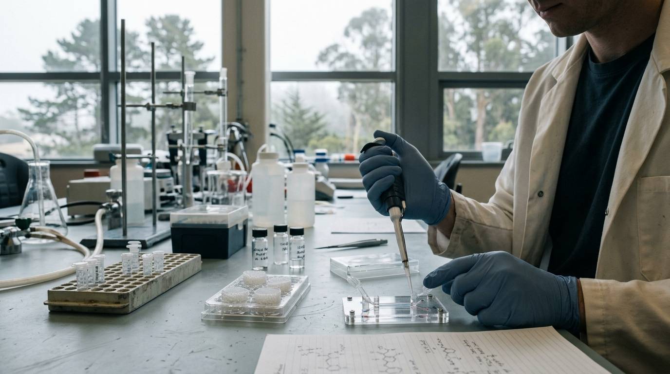
A research team sets up a preclinical screen to observe how a peptide candidate alters epithelial repair. They place the compound onto standard animal cell monolayers, observe an upregulation of signaling pathways, and prepare to publish. Yet, when other groups attempt to reproduce the signaling in primary human tissues, the biological response diverges completely.
This scenario illustrates a common challenge in regenerative research. Preclinical investigations often rely heavily on non-human animal models or immortalized cell lines. While practical, these systems frequently fail to capture human-specific cell biology.
Human-relevant platforms, including primary tissues and stem-cell-derived organoids, aim to bridge this divide. Understanding the capabilities, baseline biological variability, and functional boundaries of these advanced in vitro systems is critical when evaluating recovery, repair and regenerative research.
Defining Human-Relevant Research Models
Human-relevant models encompass experimental platforms constructed from primary human cells, adult stem cells, or induced pluripotent stem cells. Unlike two-dimensional monocultures, these systems often form complex, three-dimensional architectures that mimic the structural organization and specialized cell lineages of native organs.
In regenerative biology, investigators use these systems to study how cell signaling, extracellular cues, and prospective peptide compounds might influence tissue maintenance or repair. These platforms are research-use-only laboratory models. They are not diagnostic instruments or direct therapeutic proxies.
Researchers evaluating candidate compounds must carefully inspect whether a finding stems from an immortalized cell line, an intact rodent, or primary human tissue. The choice of system shapes what conclusions can be drawn about cellular behavior, as detailed in our guide on how to read research.
Evidence Context Across Regenerative Model Types
Experimental platforms sit on distinct tiers of biological complexity and translational relevance. Navigating this hierarchy helps prevent researchers from over-interpreting laboratory data.
Two-Dimensional In Vitro Cultures
Immortalized two-dimensional cell lines offer high throughput and low operational cost. However, they lack native three-dimensional cell polarity, multi-lineage signaling, and physiological cell-to-cell junctions. Cells cultured on flat plastic substrates often display altered gene expression patterns compared to cells in living tissue.
Animal Models
Whole-organism animal experiments capture systemic physiology, including vascular supply and systemic immune interactions. Despite these features, interspecies differences in receptor structure, metabolic processing, and tissue architecture often restrict direct extrapolation to human biology. Learning how to interpret a peptide study requires recognizing where animal biology diverges from human physiology.
Primary Three-Dimensional Human Organoids
Organoids derived from primary human tissues preserve human-specific genetics and spontaneous multi-lineage differentiation. They represent a sophisticated in vitro evidence tier. While they overcome species divergence, they remain isolated from whole-body systems such as functional immune responses and blood circulation.
Human Adult Stem-Cell Organoid Models
Evaluating epithelial regeneration requires models that accurately replicate self-renewing tissue niches. A foundational model involves human adult stem-cell-derived intestinal organoids.
In an original 2021 study, researchers established three-dimensional organoid cultures from donated human ileum and colon tissues. The investigation used tissue sourced from six adult human donors. The primary experimental goal was to determine whether adult stem cells from different individuals would display chaotic divergence or stable, reproducible behavior in laboratory culture.
The platform isolated the stem cell compartment within a supporting extracellular matrix. This environment allowed the cells to form self-organizing epithelial structures displaying crypt- and villus-like functional compartments.
Biological Pathways and Functional Evaluation
To characterize these intestinal models during differentiation, the investigators deployed several analytical modalities. These approaches evaluated whether the cultured cells possessed the functional and metabolic traits of native human tissue.
The research team tracked cellular development through:
- Gene-expression assays examining the activation of specific differentiation markers.
- Microscopy analyzing morphological architecture and multi-lineage organization.
- Hormone-secretion tests assessing endocrine cell function.
- Targeted metabolomics measuring shifts in central carbon metabolites.
Together, these readouts documented the progression of human intestinal stem cells as they differentiated into specialized epithelial lineages. By tracking central carbon metabolism alongside hormone output, the researchers verified that the cells were not merely surviving. They were actively executing tissue-specific metabolic and functional programs in vitro.
Findings on Donor Variability and Reproducibility
A recurring concern with human primary models is donor-to-donor variability. Because primary tissues retain the genetic and phenotypic diversity of individual donors, researchers often question whether experimental findings can be reproduced across multiple tissue sources.
The 2021 study addressed this question directly across its six-donor cohort. The authors reported broadly consistent differentiation pathways across the ileum- and colon-derived cultures. Metabolic shifts and developmental gene-expression markers demonstrated uniform trajectories across the tested samples.
Based on these parameters, the researchers found that donor-to-donor variability remained at a manageable level within this experimental system. This consistency suggests that primary human organoid systems can provide reproducible platforms for studying epithelial biology, provided that culture conditions and tissue-handling methods remain rigorous.
For scientists tracking peptide science and tissue response mechanisms, such baseline stability is critical. It provides an experimental baseline against which external chemical stimuli or regenerative signals can be measured.
Limitations and Experimental Unknowns
While human organoid models provide advanced human-specific biological context, their structural limits require careful consideration.
Limited Donor Sample Sizes
The primary study examined tissue from a total of six human donors. Furthermore, some individual assays tested fewer than six donors because ileum and colon cultures could not be paired for every individual. Specifically, donor 4 provided only ileal tissue, while donor 5 provided only colonic tissue.
A cohort of six donors provides an initial demonstration of technical feasibility. However, it represents an extremely narrow cross-section of broader human genetic, environmental, and demographic diversity. It cannot establish how primary cells from larger, clinically diverse populations will respond under identical conditions.
Absence of Systemic Physiology
Human organoids in standard matrix culture lack several features present in living organisms:
- Functional vasculature to deliver continuous perfusion and hydrostatic pressure.
- Circulating systemic immune cells that drive physiological inflammatory responses.
- Innervation by the peripheral nervous system.
- Direct mechanical forces, such as peristalsis or shear stress.
Because these systemic elements are absent, an effect observed in an organoid model cannot be directly assumed to occur in living human tissue. As explored in analyses of peptide research evidence levels, in vitro biological activity does not equate to clinical efficacy or safe human pharmacology.
Laboratory Verification and Testing Context
Investigating regenerative mechanisms requires high-quality analytical standards for both biological models and test compounds. In experimental biochemistry, researchers often use synthetic peptides to probe receptor pathways within organoid systems.
When sourcing materials for research use, laboratory documentation such as a Certificate of Analysis (COA) is essential. However, its exact scope must be clearly understood:
- A COA verifies the identity, molecular weight, and chromatographic purity of the specific physical sample submitted for analysis.
- Analytical purity metrics, such as high-performance liquid chromatography or mass spectrometry data, apply strictly to the vial tested.
- A COA does not prove the purity or consistency of past or future manufacturing batches.
- Chemical purity testing does not measure biological activity, cellular uptake, or metabolic stability in human tissue models.
- Laboratory reports never imply safety, clinical utility, or suitability for human administration.
Robust research standards depend on separating chemical purity verification from biological claims. Maintaining this division keeps experimental conclusions grounded in verifiable laboratory facts.
When to Revisit This Resource
Revisit this resource when designing in vitro epithelial experiments, evaluating published organoid studies, or assessing whether donor variability has been adequately addressed in human-relevant models. Rigorous model validation remains the foundation of reproducible regenerative peptide research.
Sources
Introducing / CaliLean Field Notes
A little more
perspective.
An editorial letter for curious minds. Thoughtful reading, questions worth keeping and the sources behind them.
Explore Field Notes Join the list for the first edition

