
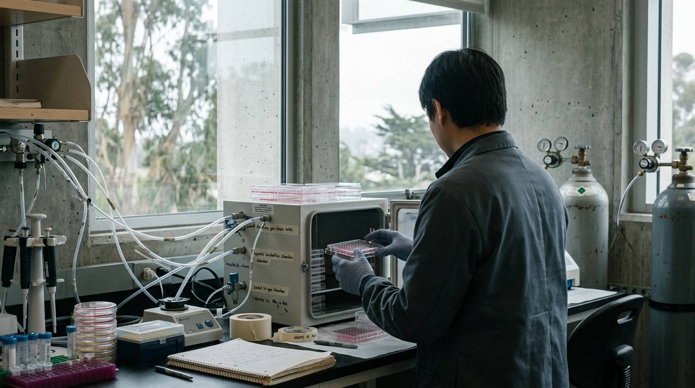
Many experimental models assume that expanding vascular density accelerates tissue closure. Preclinical data show that low oxygen environments can prompt vessel formation while stalling structural tissue reconstruction. This divergence reveals a key distinction between angiogenic signaling and functional tissue restoration. Understanding the role of oxygen tension requires looking beyond surrogate vascular markers to inspect physical measurements directly.
In regenerative biology, investigators frequently study how microenvironments dictate cellular behavior. Oxygen availability acts as a direct regulatory signal rather than a passive backdrop. Clarifying how tissues respond to hypoxic gradients helps researchers design more predictive experiments across regenerative tissue models.
Distinguish Hypoxic Responses From Structural Tissue Repair
Wound repair relies on coordinated phases of cell migration, proliferation, and extracellular matrix deposition. When researchers evaluate repair pathways, they often monitor new blood vessel growth as a benchmark of progress. However, an environment that triggers vessel sprouting does not always support rapid tissue formation. Investigating these distinct pathways prevents researchers from conflating cellular stress signals with functional tissue regeneration.
Oxygen-depleted tissue environments stabilize hypoxia-inducible factors. These factors drive the transcription of pro-angiogenic signals to restore blood supply. While this mechanism promotes localized vessel proliferation, the underlying tissue bed may lack the metabolic fuel required for cell migration. Rigorous laboratory research standards require separating these two physiological events in experimental protocols.
Evidence in this area remains grounded in animal and cell models rather than human clinical conclusions. In vitro studies demonstrate isolated cellular pathways, but they cannot reproduce the fluid dynamics of a living wound bed. Animal models bridge some of these gaps by tracking systemic responses across intact tissue layers. Researchers must interpret these findings within an established preclinical evidence hierarchy to avoid unsupported generalizations.
Track Oxygen Levels Using Direct Phosphorescence Sensors
Accurately profiling the microenvironment requires reliable physical measurements at the tissue interface. Early repair experiments often inferred oxygenation from blood flow or surrounding ambient air. Modern experimental setups measure local partial pressures directly to eliminate guesswork. Without localized verification, recorded biological effects cannot be linked to specific oxygen values.
In a controlled animal study published in PLOS ONE, investigators evaluated how surface barrier permeability altered local oxygenation. The researchers created paired, 8-millimeter full-thickness skin wounds on the backs of ddy mice aged 7 to 9 weeks. They sealed one wound with an oxygen-permeable membrane and the paired wound with an oxygen-impermeable membrane. This split design allowed direct comparison across identical genetic and systemic backgrounds.
To track tissue conditions over time, the study deployed a sensor system based on oxygen-dependent phosphorescence quenching. The Fibox 3 oxygen meter recorded real-time surface oxygen tension without consuming local oxygen during measurement. By day 7, the permeable membrane group maintained an oxygen tension equivalent to atmospheric conditions at 114.1 ± 29.8 mmHg. In contrast, the impermeable group dropped to an extremely low oxygen tension of 5.72 ± 2.99 mmHg.
Using precise sensor technology provides verifiable data that surrogate markers cannot replicate. When researchers assess experimental publications, examining these analytical tools is essential. Methodological details clarify whether observed outcomes stem from true hypoxia or incidental mechanical shielding, as outlined in guides on the critical review of experimental literature.
Evaluate Vascular Density Alongside Structural Tissue Metrics
Comparing the outcomes between the oxygenated and hypoxic mouse wounds demonstrated stark structural trade-offs. The wounds maintained under atmospheric oxygen closed significantly faster by day 7. The remaining open wound area measured 11.1 ± 1.66 percent in the oxygenated group compared to 27.6 ± 3.47 percent in the hypoxic group.
Histological analysis confirmed that oxygen availability also governed structural tissue depth. The oxygenated group formed significantly thicker granulation tissue, measuring 491.8 ± 243.2 micrometers. The hypoxic group developed a thinner granulation layer, measuring only 295.3 ± 180.9 micrometers. These metrics show that structural matrix synthesis depends heavily on sustained local oxygen levels.
Conversely, vascular analysis yielded the opposite pattern. The hypoxic wounds developed a significantly higher vascular density on day 7, reaching 0.046 ± 0.025 square millimeters per square millimeter. The oxygenated wounds recorded a vascular density of only 0.011 ± 0.008 square millimeters per square millimeter. Low oxygen tension triggered intense localized vessel growth while slowing closure and matrix production.
These findings show that vascular density alone cannot serve as an isolated metric of successful tissue repair. The mouse data prove that accelerated vessel sprouting can co-occur with delayed healing and diminished granulation thickness. Researchers must capture multiple endpoints, including closure rates and tissue volume, to obtain an accurate picture of recovery.
Account for Preclinical Model Limitations and Environmental Variables
Experimental wound models in rodents carry distinct biological constraints that limit direct translation. Murine skin heals primarily through the contraction of the panniculus carnosus muscle layer. In contrast, human skin repair relies heavily on re-epithelialization and sustained granulation formation. While the paired-wound design controls for systemic variables, the biomechanics remain specific to the ddy mouse model.
Environmental barriers also introduce physical variables beyond oxygen regulation. An impermeable membrane traps moisture, wound exudate, and metabolic byproducts along with excluding oxygen. Although sensor readings confirm the presence of profound hypoxia, the retention of fluid may also influence cellular migration. Isolating oxygen tension from moisture vapor transmission remains an ongoing challenge in preclinical wound chamber design.
Laboratory documentation and testing records must reflect these controlled parameters. A Certificate of Analysis for experimental research reagents verifies the identity and purity of a specific chemical batch. It cannot predict how a biological compound will perform under altered oxygen gradients or hypoxic stress. Researchers must carefully separate material purity reports from biological outcome models in laboratory reports.
Key Takeaways
- Low oxygen tension can stimulate angiogenic vessel sprouting while simultaneously delaying overall wound closure and granulation deposition.
- Direct measurement using phosphorescence-quenching sensors confirms that impermeable coverings create profound local hypoxia at the tissue surface.
- In paired mouse wound models, atmospheric oxygen exposure produced faster closure and thicker granulation tissue despite lower vascular density.
- Experimental designs must evaluate structural closure alongside vascular density to prevent misleading conclusions about tissue recovery.
Matching local oxygen dynamics to specific experimental models ensures that observed cellular responses reflect true biological mechanisms rather than uncontrolled environmental artifacts.
Sources
Introducing / CaliLean Field Notes
A little more
perspective.
An editorial letter for curious minds. Thoughtful reading, questions worth keeping and the sources behind them.
Explore Field Notes Join the list for the first edition

