
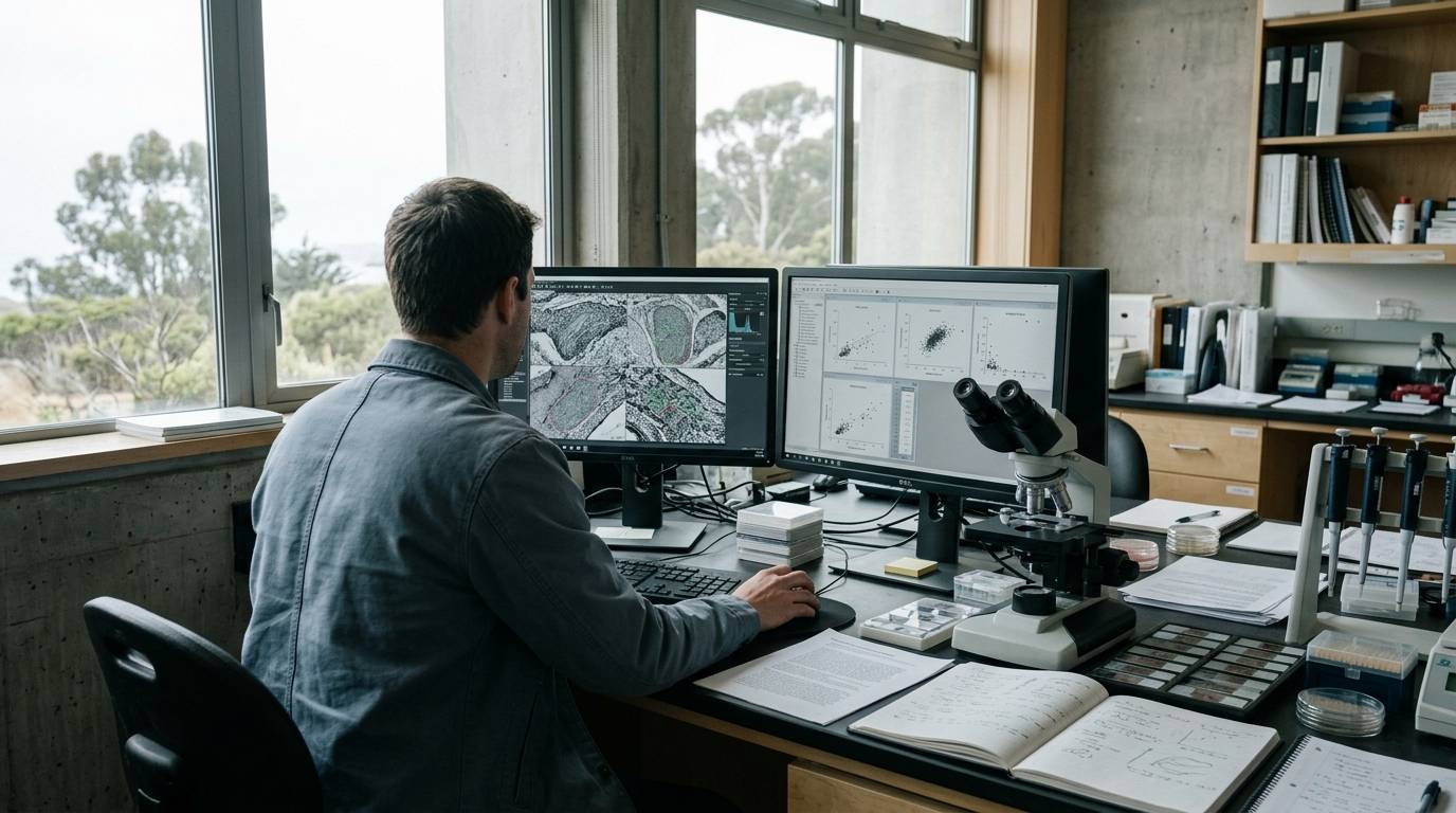
Most tissue repair assessments rely on end-point histology, assuming that a static cross-section accurately represents the dynamic recovery process. In reality, a single snapshot cannot capture the shifting vascular kinetics that occur during wound closure. Histological sampling also requires sacrificing the tissue, which eliminates the possibility of following the same animal over multiple stages of repair. Utilizing noninvasive optical modalities provides a direct window into living microvascular networks as they remodel across time.
Evaluating wound repair requires matching the imaging modality to the biological event of interest. Research teams examine cellular movement, blood vessel formation, and lymphatic vessel regeneration across different phases. Choosing an appropriate method involves balancing penetration depth, spatial resolution, and field of view. Understanding how these parameters interact helps researchers avoid drawing broad conclusions from narrow imaging data.
Track Vascular Regeneration Across Longitudinal Timelines
Preclinical imaging relies on animal research rather than controlled human data. Experimental wound assessments typically employ specialized mouse models to observe how vessels regrow after tissue injury. When evaluating these studies, researchers must understand the distinction between longitudinal observation and terminal histology. Longitudinal tracking follows individual subjects over an extended period, which reduces variability between biological samples.
Using serial measurements preserves the unperturbed wound environment throughout the healing timeline. In a study published in Light: Science and Applications, investigators utilized large-scale optoacoustic microscopy, known as LSOM, to evaluate healing skin microvasculature. The researchers imaged mouse skin vessels repeatedly during wound healing. This longitudinal approach enabled them to follow microvascular changes over time rather than relying on separate observations collected at isolated time points.
Readers evaluating preclinical wound literature should consult a guide to peptide research evidence levels to place these animal findings into an appropriate scientific context. Preclinical imaging data illuminate localized biological behaviors. However, they do not confirm how human tissues behave under similar mechanical or physiological insults.
Characterize Preclinical Study Models and Resolution Limits
The tissue model dictates which imaging tools can deliver meaningful data. In the LSOM study, researchers applied the modality to a dorsal skin full-thickness excisional wound model in female mice. Excisional models create open circular defects that heal through a mixture of wound contraction and granulation tissue formation. This physical geometry requires imaging systems that can accommodate large surface areas while still resolving individual capillary loops.
A different physical model was utilized in an investigation published in the American Journal of Pathology. In that study, researchers evaluated a mouse full-dermis thickness incision model. Incision models create narrow surgical margins rather than broad tissue voids, altering how fluid and cells migrate across the gap. The researchers tracked healing responses using noninvasive near-infrared fluorescence imaging, commonly abbreviated as NIRF.
Selecting the right imaging protocol requires researchers to critically assess the animal model, as detailed in frameworks for interpreting laboratory peptide studies. Optical penetration and skin thickness differ significantly between rodents and humans. Consequently, data gathered from dorsal mouse skin cannot be directly generalized to thicker human tissues without adjustment.
Quantify Structural Changes in Microvascular Networks
Noninvasive modalities must provide adequate spatial resolution and penetration depth to yield actionable structural data. In the female mouse excisional wound study, LSOM delivered capillary-resolution views across tissue areas spanning up to 50 mm. The optical system maintained this capillary resolution alongside a reported depth penetration of 1.5 mm. These dimensions allowed investigators to view the entire wound bed and surrounding margin without physical sectioning.
Raw images require robust computational frameworks to extract objective measurements of microvascular architecture. The LSOM researchers paired their imaging system with an automated analysis framework. This computational framework extracted several core geometric features from the regenerating vascular networks:
- Total vessel length throughout the designated region of interest.
- Vessel diameter distributions across distinct microvascular branches.
- Vessel tortuosity, which reflects the twisting nature of rapidly growing capillaries.
- Angular alignment, capturing directional sprouting toward the wound core.
Relying on standardized computational metrics reduces the subjective bias often found in manual vessel counting. Investigating these structural pathways provides clearer insights into microcirculatory remodeling. Researchers examining complex biological readouts can inspect foundational reviews of research methods to see how systematic quantification improves experimental reproducibility across different laboratories.
Differentiate Early Angiogenesis From Delayed Lymphatic Growth
Tissue repair involves multiple vascular subsystems that operate on distinct biological schedules. While blood capillaries supply oxygen and nutrients, lymphatic vessels resolve edema and clear debris from the regenerating tissue. In the mouse full-dermis incision study, researchers used longitudinal NIRF imaging to follow lymphangiogenesis over time. To validate their noninvasive fluorescence readouts, the authors confirmed the imaging observations with intravital microscopy.
The longitudinal data revealed a clear temporal difference between blood vessel growth and lymphatic vessel regeneration. Angiogenesis was observed early in the healing sequence, appearing around days 2 to 3 postincision. Conversely, NIRF-detected lymphangiogenesis emerged significantly later, appearing around days 7 to 8 after incision.
This temporal divergence illustrates why timing is critical when planning repair evaluations. Imaging an incision only during the first four days would show active angiogenesis but miss lymphatic remodeling entirely. Confirming noninvasive optical signals with secondary methods, such as intravital microscopy, ensures that fluorescent tracer signals correspond directly to true structural vessel growth.
Identify Methodological Limitations and Measurement Uncertainties
Every optical technique operates within distinct physical boundaries that introduce experimental limitations. Optical modalities experience light scattering as photons travel through dense biological tissue. In LSOM, the 1.5 mm imaging depth works well for thin rodent skin, but it cannot capture deeper subcutaneous architecture in larger organisms. Signal attenuation increases with tissue depth, which can skew automated measurements of capillary diameter in deeper dermal layers.
Fluorescence modalities present their own interpretive challenges. Tracers used in NIRF imaging depend on functional lymphatic uptake and fluid drainage kinetics. If an experimental intervention alters interstitial fluid pressure or local vascular permeability, the fluorescent signal may shift independently of true vessel growth. Researchers must verify that changes in signal intensity reflect actual vessel formation rather than simple dye pooling.
Finally, animal models have clear translational boundaries. Rodent wound healing relies heavily on panniculus carnosus contraction, whereas human wounds heal predominantly through re-epithelialization and granulation formation. Automated metrics of vessel tortuosity or angular alignment in mice cannot be interpreted as direct predictions of human scar remodeling. Researchers should evaluate imaging records with the same discipline used when examining analytical testing records in the laboratory. Each dataset reflects only the specific sample, optical parameters, and time points captured during the experiment.
Key Takeaways
- Longitudinal imaging enables researchers to follow the same living subject over time, avoiding the destructive artifacts of terminal histology.
- In female mouse excisional wound models, LSOM demonstrated capillary-resolution imaging across areas up to 50 mm with a 1.5 mm depth penetration.
- Automated analytical tools allow objective measurement of microvascular structural features, including vessel length, diameter, tortuosity, and angular alignment.
- Blood vessel and lymphatic vessel formation follow distinct timelines, with angiogenesis appearing around days 2 to 3 and lymphangiogenesis emerging around days 7 to 8 in mouse incision models.
- Imaging observations gathered via noninvasive modalities benefit from secondary validation methods, such as intravital microscopy, to confirm underlying vascular structures.
- Preclinical optical data must not be conflated with clinical human outcomes due to differences in tissue thickness, contraction kinetics, and optical penetration limits.
Rigorous evaluation of tissue repair requires aligning the imaging modality, temporal sampling window, and analytical framework with the physical limits of the biological model.
Sources
Introducing / CaliLean Field Notes
A little more
perspective.
An editorial letter for curious minds. Thoughtful reading, questions worth keeping and the sources behind them.
Explore Field Notes Join the list for the first edition

