
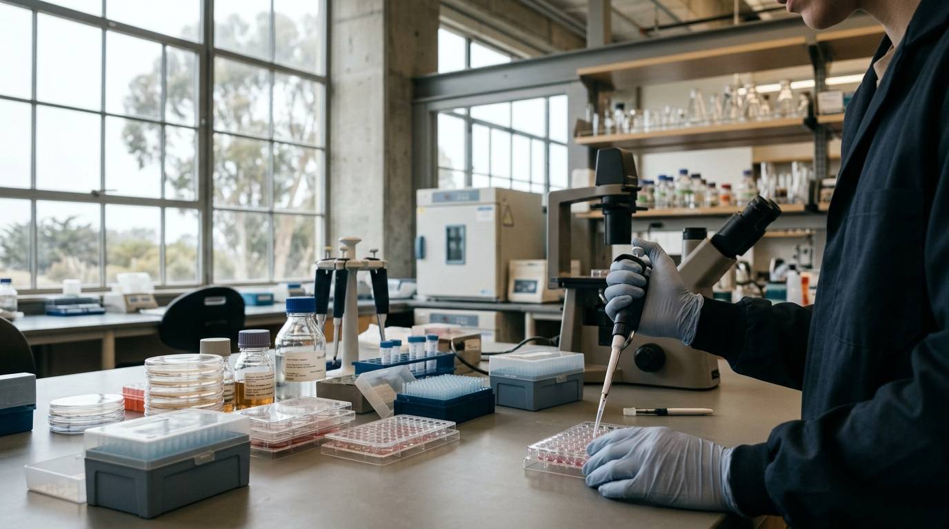
Wound healing in the lung does not begin with cell division. Instead, airway epithelial cells first dismantle their mature identities and migrate across the wound bed before proliferation occurs. Understanding how the respiratory epithelium restores its physical barrier requires close attention to experimental architecture. This guide evaluates how in vitro models measure physical closure, marker expression, and immune interactions during respiratory repair.
Examine the Mechanics of Airway Barrier Restoration
Evaluating pulmonary epithelial repair requires separating barrier integrity from basic wound closure. While visual closure can appear complete under microscopy, functional restoration depends on the formation of tight junctions. Researchers track this physical sealing through transepithelial electrical resistance, often abbreviated as TEER. Measuring TEER provides an objective readout of ion permeability across the epithelial layer.
Primary cell cultures show that barrier recovery correlates closely with the scale of physical disruption. In controlled scratch assays, transepithelial electrical resistance returned fully to baseline within 40 hours following small mechanical injuries. Larger injuries failed to achieve complete sealing within a similar timeframe. Even after 44 hours, larger wound areas recovered to only 70 to 80 percent of control resistance values.
Physical barrier restoration does not immediately follow cellular repopulation of the wounded surface. Cells must establish polarized apical and basolateral domains while restoring continuous junctional complexes. Without these restored junctions, the epithelial barrier remains leaky despite confluent visual coverage. Research teams must evaluate experimental models of cellular activity by inspecting electrophysiological measurements alongside structural coverage.
Distinguish Cell Culture Evidence from Organ Function
The primary evidence for respiratory barrier restoration comes from in vitro cell culture models. Air-liquid interface cultures, or ALI systems, represent the current standard for primary human small airway epithelial cells. By exposing the apical surface of the cells to air, these platforms induce mucociliary differentiation. Ciliated cells, club cells, and goblet cells form a pseudostratified layer that mirrors human airway tissue.
Cell culture evidence operates under distinct biological boundaries compared to intact lung tissue. Isolated epithelial sheets lack capillary perfusion, mechanical ventilation pressures, and normal lymphatic drainage. Preclinical cell findings reveal isolated cellular behaviors, but they cannot represent whole organ physiology. Understanding these distinctions is critical when assessing the translational risk of preclinical data.
The baseline evidence level for these detailed scratch models remains strictly in vitro. The physical forces applied by mechanical scratch tools simulate acute trauma rather than chronic airway disease. Furthermore, the absence of an underlying vascular matrix simplifies the repair environment. Readers must treat these systems as mechanistic testbeds rather than direct clinical proxies.
Track Epithelial Phenotype and Junction Remodeling
During mechanical injury resolution in air-liquid interface systems, cellular repair proceeds through an ordered sequence. Differentiated apical cells at the wound edge rapidly de-differentiate. These altered cells migrate directly into the denuded space. Only after this initial migratory phase do apical cells behind the leading edge begin active proliferation.
This migratory phase coincides with significant changes in molecular markers between 24 and 72 hours post-injury. The repair process involves a sharp reduction in tight junction molecules:
- Claudin-3 (CLDN3) expression decreases during active cell migration.
- Occludin (OCLN) levels drop as junctional complexes temporarily disassemble.
Markers of mature airway cell types experience a simultaneous reduction:
- FOXJ1, a primary transcription factor for ciliated cells, declines rapidly.
- SCGB1A1, characteristic of secretory club cells, exhibits marked down-regulation.
- TFF3, an indicator of columnar epithelial cells, shows reduced expression.
- ACTA2, coding for alpha-smooth muscle actin, also decreases during this window.
At the leading edge of the wound, cells up-regulate focal adhesion kinase within 24 hours. This kinase activation supports the cellular motility required to traverse the exposed surface. In parallel, injured cells increase both messenger RNA and protein levels of fibronectin. Fibronectin appears intracellularly at the wound edge before depositing into the extracellular matrix to support migration.
Evaluate Macrophage Crosstalk in Wound Repair
Airway repair takes place within a dynamic immune environment. To examine this interaction, researchers have paired primary airway epithelial cells with human monocyte-derived macrophages. Monocytes can be differentiated in the laboratory into M1 phenotypes using GM-CSF or M2 phenotypes using M-CSF. Introducing these immune populations into air-liquid interface cultures reveals distinct regulatory signals.
The presence of immune cells directly influences epithelial differentiation states. Laboratory experiments demonstrate that both M1 and M2 macrophage subtypes promote epithelial de-differentiation. In addition, M1 macrophages stimulate increased epithelial expression of focal adhesion kinase. This response indicates that inflammatory signals actively participate in priming epithelial cells for migration.
Subsets of macrophages exert divergent effects on matrix production and cell growth. While general phenotypes encourage cell mobilization, M2a macrophage subsets introduce inhibitory signaling. Specifically, M2a macrophages suppress both epithelial proliferation and fibronectin production. This suppression demonstrates that immune cells do not simply accelerate healing, but instead govern distinct checkpoints.
Recognize Experimental Constraints and Model Discrepancies
Laboratory systems carry unavoidable technical and biological limitations. In primary epithelial scratch assays, the integrity of the underlying collagen-coated membrane is paramount. Experimental findings show that mechanical damage to this underlying collagen coat severely impairs the initial migratory process. While cells can eventually span damaged membranes when opposing edges converge, substrate integrity remains a confounding variable.
Technical limitations also constrain immune co-culture observations. In published primary cell systems, researchers could not determine whether macrophage subtypes altered the actual rate of wound closure. While molecular markers shift in response to macrophages, measuring overall closure kinetics across complex multi-cell platforms remains technically challenging.
The mode of tissue injury creates notable discrepancies in cellular behavior across the literature:
- Mechanical scratch injuries trigger migration driven primarily by de-differentiated apical cells.
- Chemical injury models frequently recruit basal progenitor cells to drive barrier restoration.
- Oxidative damage models also document a primary role for basal stem cells rather than apical sheet migration.
Researchers cannot assume that repair pathways identified through mechanical scratches apply equally to toxin exposure or oxidative stress. Every physical model isolates only a small subset of the physiological mechanisms active in human respiratory tissue.
Interpret Laboratory Data Without Clinical Extrapolation
Data generated from primary cell platforms must be interpreted with analytical discipline. Biochemical characterizations, cell survival assays, and peptide exposure studies reveal specific cellular behaviors under static conditions. These observations do not provide human safety data, metabolic profiles, or therapeutic guidelines. Analytical purity records and verified laboratory test methods clarify compound quality, but they cannot predict complex tissue biology.
When reviewing compound analyses or evaluating laboratory testing records, the scope remains strictly limited to the tested sample. A laboratory certificate confirms the chemical identity or purity of a single batch under specified conditions. It does not establish biological efficacy or clinical tolerability in an intact lung. Responsible analysis demands that investigators separate physical measurements from broad biological conclusions.
Key Takeaways
- Barrier restoration is injury-dependent, with smaller scratches recovering normal electrical resistance within 40 hours, while larger wounds reach only 70 to 80 percent of baseline after 44 hours.
- Airway repair begins with the de-differentiation and migration of apical cells, while cellular proliferation occurs secondary to initial coverage.
- Active closure temporarily down-regulates tight junction markers (CLDN3, OCLN) and lineage markers (FOXJ1, SCGB1A1, TFF3, ACTA2) while up-regulating focal adhesion kinase and fibronectin.
- M1 and M2 macrophages encourage epithelial de-differentiation, while M2a phenotypes specifically suppress proliferation and fibronectin accumulation.
- Substrate damage directly impairs cell migration, and mechanical scratch responses differ substantially from chemical or oxidative repair mechanisms.
Rigorous pulmonary research requires evaluating in vitro models on their own merits, acknowledging their boundaries, and avoiding premature clinical conclusions.
Sources
Introducing / CaliLean Field Notes
A little more
perspective.
An editorial letter for curious minds. Thoughtful reading, questions worth keeping and the sources behind them.
Explore Field Notes Join the list for the first edition

