
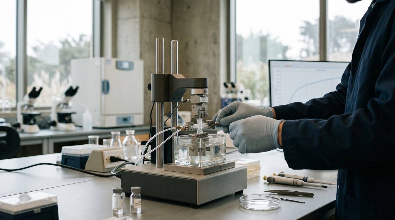
High cellularity and organized matrix deposition under a microscope do not always guarantee that repaired tissue can bear real physiological loads. Many investigators assume that restored structural appearance immediately equates to restored physical strength. In musculoskeletal regeneration research, mechanical testing evaluates whether repaired tissues regain physical load-bearing properties. Evaluating these physical responses under force clarifies whether a healing matrix has regained functional integrity or remains vulnerable to mechanical failure.
To interpret these investigations accurately, readers must review how to read preclinical research and understand the physical benchmarks used in laboratory settings.
Differentiate Structural Properties From Material Properties
Biomechanical evaluation requires a clear distinction between whole-structure responses and intrinsic tissue behavior. According to a review of vertebrate and mammalian tendon models published in the PMC literature, researchers evaluate two distinct categories of physical measurements during tensile experiments.
Structural properties reflect the response of the entire specimen as a complete physical unit. These parameters are measured as load-to-failure and overall deformation during tensile testing. Because total tissue volume, thickness, and cross-sectional area directly influence these values, structural metrics alone cannot isolate the underlying matrix quality from specimen geometry.
Material properties describe the intrinsic quality of the matrix itself regardless of total specimen size. These parameters normalize the physical forces against the dimensions of the sample. Material properties include measures of tissue stress, strain, and elastic modulus. Calculating these values allows investigators to assess whether the newly organized matrix matches healthy native tissue at the compositional level.
Understanding this division prevents misleading conclusions in regenerative models. A large, disorganized scar mass might withstand a high absolute load simply due to bulk volume. However, its material modulus and stress capacity may remain substantially inferior to uninjured native tissue.
Correlate Histological Structure With Functional Integrity
Microscopic evaluation and physical load testing offer distinct windows into the healing process. While physical performance often diverges from visual observation, clear structural alignment can align with mechanical stability in specific biological contexts.
In an original rat Achilles tendon repair study published by researchers in PMC, histology scores were plotted directly against biomechanical test outcomes across healing tendons. The investigation reported a strong linear correlation between histological scores and biomechanical characteristics. In that specific rodent model, organized cellular and collagen architecture closely mirrored the recovery of tensile strength.
Even so, researchers cannot assume this correlation holds universally across every animal model or tissue architecture. Fibrotic repair tissue often looks dense and organized under light microscopy, yet it may fail to replicate native collagen cross-linking. Combining mechanical evaluations with histological staging remains essential to verify true regenerative recovery. Investigators reviewing recovery and repair peptide research should always inspect whether studies test mechanical integrity or merely report cellular appearance.
Control Specimen Geometry and Grip Interfaces
Applying standard tensile protocols to biological soft tissues presents severe methodological hurdles. Tensile testing is an essential experiment to assess the mechanical integrity of musculoskeletal soft tissues. However, standardized test methods have not been fully established to ensure quality and reproducibility across laboratories.
Specimen preparation significantly alters measured outcomes. In an in vitro study evaluating bovine meniscus soft tissue coupons, researchers investigated how standard ASTM coupon geometries altered mechanical measurements. The study showed that bovine meniscus tensile results differed significantly based purely on coupon shape.
Dumbbell-shaped coupons yielded greater measured ultimate strength and modulus compared to straight rectangular coupons. The geometry of the specimen also dictated where the tissue ruptured during tensile pulling:
- Dumbbell coupons cut according to ASTM standards for elastomers achieved an 80% rate of midsubstance failures.
- Dumbbell coupons cut according to ASTM standards for plastics produced a 60% rate of midsubstance failures.
- Straight coupons prepared according to ASTM standards for fiber-reinforced composites dropped to a 20% rate of midsubstance failures.
When specimens fail at the clamping grips rather than within the midsubstance gauge length, the recorded failure loads underestimate true tissue strength. Stress concentrations near clamping fixtures often induce premature failure. Adopting standardized specimen geometries that promote midsubstance failure is necessary to capture authentic tissue capability.
Diagnose Sources of Experimental Variability
Musculoskeletal soft tissues are inherently anisotropic, hydrated, and viscoelastic. These biological realities introduce variability that standard engineering protocols struggle to accommodate without strict controls.
Variations in tissue hydration during testing dramatically skew stress-strain curves. Dehydration increases apparent stiffness and brittle failure modes, while over-hydration can alter cross-sectional measurements. Cross-sectional area measurement itself remains a notorious source of calculation error. Because soft tissues deform under contact calipers, non-contact optical methods are frequently required to prevent artificial distortion of stress and modulus values.
Preconditioning protocols also shape recorded outcomes. Soft tissues exhibit hysteresis and load-history dependence. Subjecting specimens to cyclical loading before running a failure test aligns collagen bundles and establishes a stable reference baseline. Studies that omit preconditioning or fail to report strain rates create data that cannot be compared reliably against external literature.
Adhering to rigorous research standards requires documenting temperature, bath conditions, clamping pressure, and strain velocity. Without these precise reporting details, subtle shifts in mechanical outcomes might reflect benchtop artifacts rather than true biological regeneration.
Inspect Laboratory Reports and Analytical Scope
Translating tissue-engineering concepts into reliable benchwork requires clean, verified experimental reagents. When working with research peptides or biomaterial scaffolds, laboratories confirm chemical identity and purity before initiating complex mechanical trials.
Analytical methods such as high-performance liquid chromatography and mass spectrometry establish the chemical integrity of experimental compounds. These details are reported on laboratory certificates of analysis. However, researchers must understand the clear boundaries of these documents.
An analytical report confirms only the identity and purity of the specific sample submitted to the testing equipment. It does not establish how that material will behave inside a biological matrix, nor does it guarantee mechanical performance in vivo. Verifying compound purity ensures that experimental variability stems from biological responses rather than uncharacterized chemical contaminants.
Key Takeaways
- Structural properties measure overall physical limits like load-to-failure and deformation, whereas material properties measure intrinsic matrix metrics including stress, strain, and elastic modulus.
- An original rat Achilles tendon study demonstrated a strong linear correlation between histological scores and biomechanical recovery, but this relationship must be verified across distinct tissue types.
- Specimen geometry substantially alters test outcomes, as demonstrated by bovine meniscus coupons where dumbbell shapes showed higher measured strength and superior midsubstance failure rates compared to straight strips.
- Standardized testing guidelines for soft tissues remain limited, making clear reporting of hydration, strain rates, and clamping methods essential for reproducibility.
- Analytical testing and documentation verify compound purity for the specific sample tested, but they do not predict mechanical efficacy in living tissue systems.
Mechanical testing remains the definitive standard for proving that regenerated soft tissues can handle physical force rather than simply mimicking native tissue under a microscope.
Sources
Introducing / CaliLean Field Notes
A little more
perspective.
An editorial letter for curious minds. Thoughtful reading, questions worth keeping and the sources behind them.
Explore Field Notes Join the list for the first edition

