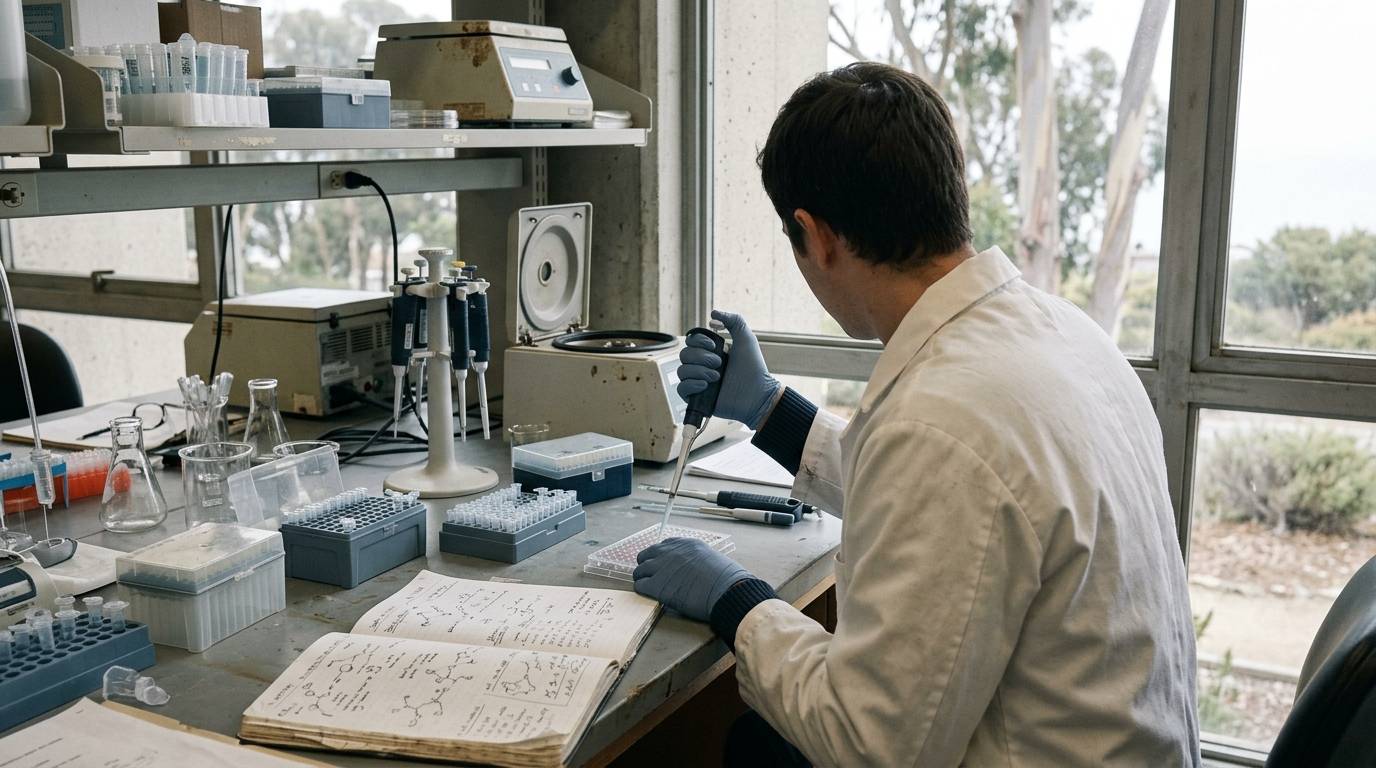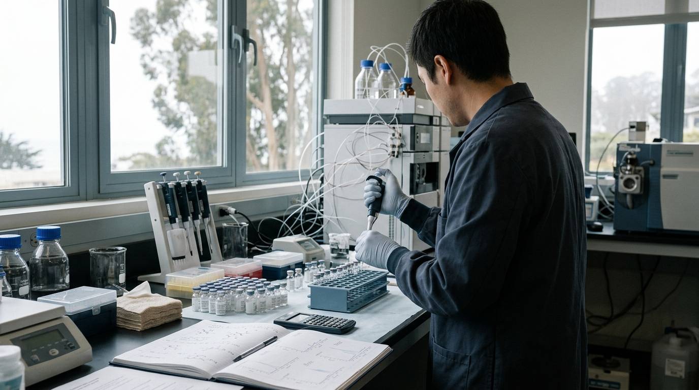
A single high percentage on an analytical readout does not prove that a peptide sample is chemically pure. In routine analytical practice, a high-performance liquid chromatography chromatogram showing a ninety-eight percent peak area can obscure substantial co-eluting species or unobserved contaminants. Establishing chemical identity and purity requires an understanding of what each laboratory technique measures and what it overlooks. This guide examines the analytical frameworks used to characterize synthetic peptides, focusing on the evidence generated by liquid chromatography and high-resolution mass spectrometry.
Evaluating synthetic peptide quality depends on recognizing the boundaries of specific laboratory methods. Purity is not a single, universally measurable property. Instead, purity reflects the absence of specific classes of contaminants as defined by a chosen test. A method designed to separate molecules based on hydrophobicity will fail to capture impurities that do not interact with the column matrix. Addressing these challenges requires orthogonal testing strategies and transparent reporting of analytical limits.
Distinguish Study Models and Evidence Levels
Analytical peptide characterization operates strictly within an in vitro laboratory context. The published data evaluating chromatographic and mass spectrometric performance rely on synthetic peptide reference materials rather than biological systems. These experiments evaluate chemical stability, chromatographic resolution, and ionization behavior in controlled instrument environments.
Controlled laboratory studies frequently employ synthetic peptide fragments to benchmark analytical performance. For instance, researchers characterized the analytical behavior of a 21-residue synthetic peptide corresponding to an S-carbamidomethylated tryptic fragment of human serum albumin, designated as cam-it3. In another investigation, researchers analyzed synthetic human C-peptide materials to develop impurity profiling workflows. Neither of these investigations evaluated biological efficacy, cellular responses, or clinical outcomes in human populations.
Conflating analytical method development with biological performance undermines scientific rigor. A method that successfully isolates a target peptide in a clean solvent mixture cannot predict how that peptide functions in vivo. The purpose of these studies is solely to refine measurement accuracy, quantify known synthetic artifacts, and establish chemical characterization protocols. Understanding these distinctions helps researchers navigate peptide research evidence standards without assuming that physical characterization translates directly to physiological activity.
Challenge Common Peak-Area Assumptions in Liquid Chromatography
A standard method for estimating peptide purity relies on high-performance liquid chromatography coupled with ultraviolet detection, often referred to as HPLC-UV. In this configuration, the analyst integrates the detector response over time and expresses the target peak area as a percentage of the total detected peak area. While widespread, this approach relies on a critical theoretical assumption. It assumes that the peptide of interest, any related peptide impurities, and all non-peptide impurities generate an ultraviolet response that is directly proportional to their mass.
Original laboratory research demonstrates that this assumption is not always valid. In an investigation examining synthetic peptides, including the 21-residue cam-it3 peptide and it3, researchers cautioned against treating UV peak area percentages as direct mass-basis purity corrections. Different peptide sequences contain varying numbers of chromophores, such as aromatic amino acids and peptide bonds. Consequently, truncation products or deletion sequences can display radically different extinction coefficients compared to the full-length target.
When an impurity produces a weaker UV signal per unit of mass than the parent sequence, integrating UV peak areas underestimates the impurity content. Conversely, an impurity with strong absorbance can artificially depress the apparent purity of the target molecule. Non-peptide impurities, including residual counterions, synthesis scavengers, and moisture, often fail to absorb UV light at standard detection wavelengths entirely. Relying solely on HPLC-UV peak area percentages can therefore overestimate the mass fraction of the target peptide within a dry material sample.
For researchers consulting a peptide certificate of analysis, recognizing these UV response differences is essential. An area-percent value reflects only the relative absorbance of detected, light-absorbing components under specific chromatographic conditions. It does not provide an absolute mass-fraction measurement of total chemical content.
Identify Structurally Related Impurities with High-Resolution Methods
Because standard UV detection cannot resolve closely related peptide variants, advanced laboratory protocols employ orthogonal separation and detection techniques. Liquid chromatography coupled to high-resolution mass spectrometry, or LC-HRMS, provides both retention time separation and accurate mass-to-charge measurements. This approach allows laboratories to identify impurities that share near-identical chromatographic retention times with the target molecule.
Synthetic peptide manufacturing processes routinely generate structurally related impurities through incomplete coupling, side-chain deprotection failures, or oxidation. In a published laboratory study, researchers developed LC-HRMS methods to evaluate synthetic human C-peptide study material. Their targeted analytical characterization identified, confirmed, and quantified more than 65 distinct impurities within the study material.
These findings illustrate the depth of chemical complexity present in synthetic peptide preparations. A standard HPLC run with broad integration parameters would merge many of these structurally related variants into the baseline or the primary peak shoulders. By contrast, high-resolution mass spectrometry distinguishes subtle molecular differences, including:
- Deamidation events that alter peptide mass by less than one Dalton.
- Truncation sequences missing a single hydrophobic amino acid.
- Diastereomers and insertion sequences resulting from incomplete solid-phase synthesis cycles.
- Oxidation variants formed during cleavage, handling, or storage.
Applying high-resolution instruments demonstrates that targeted characterization reveals components beyond a simple target-peak readout. Mass spectrometry provides critical sequence confirmation by matching observed monoisotopic masses against theoretical fragmentation patterns. However, mass spectrometry also introduces specific detection biases, as ionization efficiency varies significantly across diverse peptide structures. For more technical background on instrument workflows, review our guide to understanding analytical methods in laboratory research.
Assess Method Limitations and Documentation Scope
Every analytical instrument operates within defined limits of detection, linear dynamic ranges, and chemical selectivity. Liquid chromatography separates molecules based on partition coefficients, meaning that strongly retained or completely unretained contaminants may never reach the detector. Mass spectrometers depend on analyte ionization, so neutral species or compounds that suppress electrospray ionization remain undetectable in standard workflows.
These technical boundaries mean that no single analytical run can establish complete chemical purity. An HPLC-UV chromatogram confirms the optical uniformity of detected peaks at a selected wavelength. An LC-HRMS spectrum confirms the presence of molecules with specific mass-to-charge ratios. Neither test inherently quantifies total net peptide content, residual salts, or dissolved water.
Furthermore, laboratory documentation must be interpreted within its original context. An analytical report or Certificate of Analysis applies strictly to the specific aliquot submitted for instrument testing. A report confirms that the tested sample met specific instrumental parameters under documented chromatographic conditions on a particular date. It does not establish that every aliquot in a production lot is identical.
Analytical documentation provides an objective record of test results, but it cannot establish biological properties. A high purity readout from an LC-HRMS system does not confirm biological stability or safety. Laboratory reports document chemical metrics for research materials, supporting reproducibility across experimental designs. Researchers can consult the broader peptide science resources to learn how different analytical metrics integrate into rigorous laboratory studies.
Key Takeaways
- HPLC-UV area percentage assumes that all components absorb light proportionally to their mass, which is often invalid.
- Non-absorbing impurities, including salts and moisture, are invisible to standard UV detection protocols.
- High-resolution mass spectrometry can resolve dozens of structurally related impurities that co-elute with the target peptide.
- A laboratory test report establishes analytical values only for the specific sample tested under documented instrument conditions.
Understanding the direct assumptions and technical boundaries of analytical tools ensures that peptide research data are interpreted with appropriate scientific restraint.
Sources
Introducing / CaliLean Field Notes
A little more
perspective.
An editorial letter for curious minds. Thoughtful reading, questions worth keeping and the sources behind them.
Explore Field Notes Join the list for the first edition