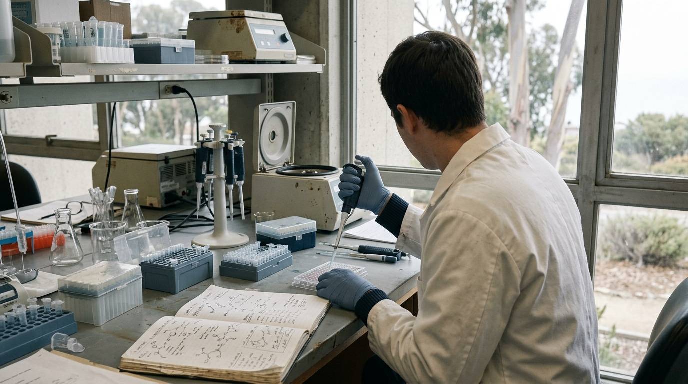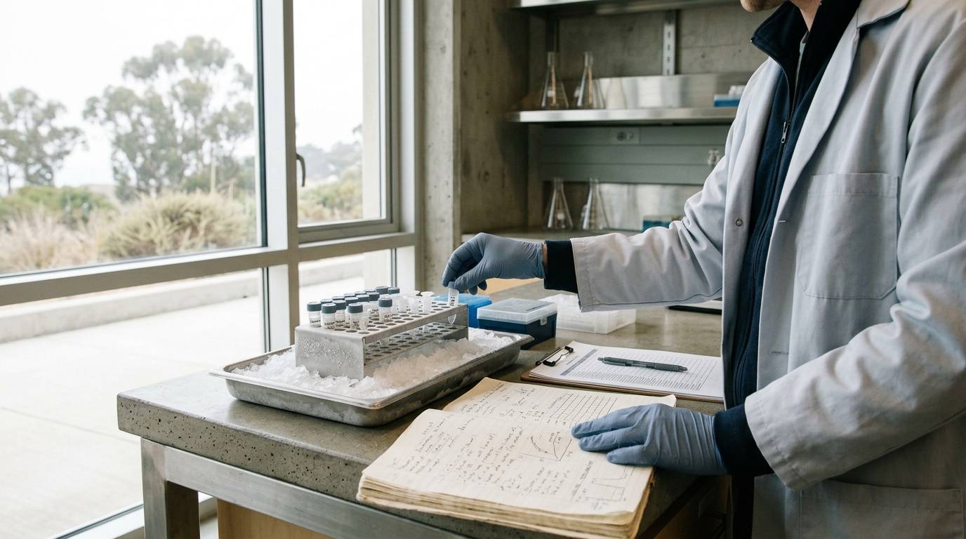
Analytical assays often produce precise numbers that lead researchers to assume their target peptide remained intact throughout processing. In metabolic research, that assumption is frequently incorrect. Peptides can undergo rapid cleavage within minutes of blood collection, leaving behind fragments that standard assays cannot always differentiate from intact molecules. Understanding how collection tubes, temperature, and analytical choices influence sample integrity is essential for generating reliable laboratory measurements.
This resource examines how pre-analytical handling alters measured concentrations of key metabolic peptides. It details the enzymatic pathways responsible for rapid degradation, highlights discrepancies between mass spectrometry and immunoassays, and reviews stability limits in common specimen matrices.
Distinguish Specimen Models and Analytical Evidence
Research into metabolic peptide stability relies primarily on in vitro experiments using ex vivo human blood specimens. These laboratory evaluations expose whole blood, plasma, or serum to controlled handling conditions to trace molecular degradation over time. They provide direct insight into pre-analytical chemistry, but they do not describe therapeutic actions in living subjects.
Because stability profiles differ markedly across molecules, laboratory conclusions from one compound cannot be applied universally. Researchers must inspect the specific experimental conditions, matrix additives, and detection systems used in each investigation. Research published in the peptide fundamentals and biology literature shows that specimen stability depends on sequence-specific enzymatic susceptibility rather than molecular weight alone.
The findings discussed below come from three distinct in vitro laboratory evaluations. One investigation analyzed human blood specimens spiked with incretin and metabolic peptides using mass spectrometry alongside immunoassays. A second study evaluated spiked plasma specimens from a pool of 20 healthy volunteers to test temperature and freeze-thaw parameters. A third study assessed endogenous insulin and C-peptide levels in whole blood and serum specimens collected from five healthy volunteers.
Track Enzymatic Breakdown and Assay Divergence
Enzymatic degradation begins immediately when biological samples are collected without appropriate inhibitors. In human blood specimens, dipeptidyl peptidase-4 (DPP-IV) cleaves the two N-terminal amino acids from glucagon-like peptide-1 (GLP-1) and glucose-dependent insulinotropic polypeptide (GIP). In non-stabilized human blood samples, researchers observed that these N-terminal fragments and subsequent breakdown products accumulated rapidly as the specimens stood at bench conditions.
The choice of detection platform plays a critical role in whether this breakdown is noticed. In human blood studies monitoring GLP-1, GIP, glucagon, and oxyntomodulin via matrix-assisted laser desorption ionization time-of-flight mass spectrometry (MALDI-TOF MS) and antibody-based assays, significant measurement discrepancies appeared. While the antibody-based assay for GLP-1 successfully tracked intact, active GLP-1, the antibody-based assays for GIP, glucagon, and oxyntomodulin failed to reflect the progressive degradation detected by mass spectrometry.
This analytical blind spot occurs because antibodies frequently bind to epitopes that remain intact on cleaved peptide fragments. As a result, an immunoassay can report stable concentrations even as the bioactive parent peptide disappears from the sample. Researchers relying solely on antibody detection may therefore overestimate analyte concentrations in degraded specimens. Documenting the specific analytical methods used to verify sample integrity is necessary to avoid misinterpreting metabolic data.
Control Collection Tubes and Thermal Variables
Thermal management during processing dictates how much intact peptide survives prior to testing. In pooled human plasma containing a DPP-IV inhibitor and aprotinin, holding samples at room temperature for longer than one hour significantly impaired the recovery of both GLP-1 and glucagon. Conversely, holding the plasma specimens on ice for up to three hours maintained peptide recovery without measurable loss.
Freeze-thaw behavior also presents distinct challenges depending on the target analyte. In that same pooled plasma study, subjecting specimens to up to three freeze-thaw cycles did not alter peptide recovery. Long-term frozen storage revealed a striking divergence between analytes. GLP-1 concentrations remained stable in frozen plasma for at least one year, but glucagon recovery dropped by nearly 50% immediately upon freezing compared with fresh analysis, regardless of total storage time.
Sample collection tubes further dictate analytical stability. An in vitro evaluation using blood from five healthy volunteers compared potassium-EDTA tubes with serum-gel tubes. Insulin concentrations remained stable for 24 hours at room temperature in both centrifuged plasma and uncentrifuged whole blood collected in EDTA tubes. In contrast, insulin levels in serum-gel tubes declined significantly by 24 hours, dropping to 66% of baseline in centrifuged serum and 76% of baseline in uncentrifuged specimens.
In that same study, C-peptide demonstrated stability across multiple handling conditions. However, C-peptide levels declined significantly when left in uncentrifuged serum-gel tubes. Implementing proper stability and storage protocols tailored to each specific peptide and matrix prevents these pre-analytical artifacts from corrupting study datasets.
Evaluate Documentation and Methodological Limits
Published stability experiments carry notable methodological limitations that researchers must account for during experiment design. In vitro spiking studies often use synthetic peptides at concentrations that differ from physiological baseline levels. Spiked peptides may not reflect the exact protein-binding dynamics or degradation rates found in endogenous circulation.
Cohort sizes in stability literature are frequently small. A study relying on pooled plasma from 20 individuals or whole blood from five subjects cannot account for patient-level metabolic variability. Differences in endogenous protease activity, kidney function, or anticoagulant mixing can introduce variance not captured in small laboratory cohorts.
Furthermore, laboratory certificates cannot compensate for handling errors that occur after receipt. When reviewing technical specifications or interpreting a peptide certificate of analysis, researchers must understand that chromatographic purity applies only to the tested dry material. A Certificate of Analysis does not guarantee stability once the peptide enters a biological matrix. Reference documents such as peptide identity and content standards outline lot verification, but they do not establish stability under variable laboratory handling.
Diagnose Pre-Analytical Vulnerabilities
To preserve specimen validity, laboratory workflows should account for specific pre-analytical vulnerabilities:
- Rapid enzyme action: DPP-IV quickly cleaves N-terminal residues from incretin peptides unless specific inhibitors are introduced at venipuncture.
- Cross-reactive fragments: Immunoassays for peptides like GIP and glucagon can bind truncated fragments, giving an illusion of stability where degradation has occurred.
- Temperature sensitivity: Keeping plasma at room temperature beyond 60 minutes impairs recovery, whereas ice baths preserve integrity for up to three hours.
- Matrix incompatibility: Serum-gel tubes lead to significant losses in insulin and C-peptide over 24 hours compared to EDTA tubes.
- Freeze damage: Certain peptides such as glucagon experience substantial recovery drops following a single freeze event.
Key Takeaways
- Pre-analytical degradation can occur within minutes, altering target peptide structure before analytical measurement takes place.
- DPP-IV cleaves two N-terminal amino acids from GLP-1 and GIP, generating inactive metabolites in non-stabilized blood.
- Immunoassays for GIP, glucagon, and oxyntomodulin can fail to detect progressive degradation that is clearly visible under mass spectrometry.
- Room-temperature bench handling beyond one hour reduces peptide recovery, while ice baths maintain sample stability for up to three hours.
- Freezing plasma reduces glucagon recovery by roughly 50% compared to fresh analysis, whereas GLP-1 remains stable frozen for at least one year.
- Potassium-EDTA preserves insulin stability at room temperature for 24 hours, whereas serum-gel tubes permit significant degradation.
Rigorous pre-analytical documentation ensures that metabolic measurements reflect genuine biological differences rather than variable sample handling.
Sources
Introducing / CaliLean Field Notes
A little more
perspective.
An editorial letter for curious minds. Thoughtful reading, questions worth keeping and the sources behind them.
Explore Field Notes Join the list for the first edition