
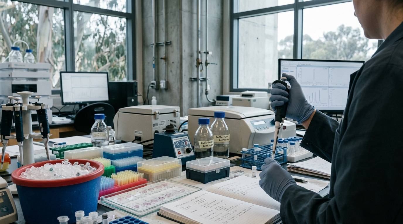
Researchers frequently search for why a peptide candidate demonstrating nanomolar affinity in a cell culture screen fails to elicit neurochemical responses in an intact animal model. The direct answer is that nominal concentration and administered dose do not dictate biological activity within the central nervous system. Between systemic administration and receptor activation lie enzymatic barriers, vascular constraints, and tissue clearance pathways that dramatically alter the physical molecule.
A peptide may degrade before reaching cerebral vessels. It may clear rapidly through renal filtration or become trapped within capillary endothelial membranes. Even when parenchymal entry occurs, rapid tissue binding or enzymatic inactivation can prevent interaction with intended neurobiological targets.
This guide examines how stability, metabolism, and pharmacokinetics govern peptide behavior in laboratory neuroscience. We evaluate the core pathways of enzymatic processing, blood-brain barrier interfaces, and intra-brain spatial dynamics. By framing peptide action through local molecular exposure rather than gross dosing, researchers can build reproducible protocols and interpret laboratory observations accurately.
For additional frameworks on evaluating preclinical publications, see our guide on how to read research and avoid common translational assumptions.
Pharmacokinetic Frameworks for Central Nervous System Peptides
Standard pharmacokinetic models use the classical absorption, distribution, metabolism, and excretion framework. For neuroscience investigations, this general framework must expand to account for complex central nervous system boundaries. A compound must first maintain adequate systemic circulation to reach the cerebral vascular network.
Once blood carries a peptide to the brain microvasculature, it encounters the endothelial lining of the blood-brain barrier. Crossing this boundary does not guarantee widespread parenchyma distribution. The peptide must escape clearance back into the systemic circulation, avoid local enzymatic degradation, and diffuse through the brain extracellular fluid.
Distribution within the central nervous system comprises distinct spatial compartments. These compartments include the cerebral vasculature, the vascular endothelial cells, the extracellular interstitial fluid, the cerebrospinal fluid, and intracellular spaces. Concentrations in one compartment rarely equal concentrations in another.
- Systemic Circulation
- (Enzymatic Cleavage & Renal Clearance)
- Cerebral Microvasculature
- (Endothelial Transport vs. Efflux)
- Brain Extracellular Fluid (Interstitial Space)
- (Diffusion vs. Extracellular Peptidases)
- Neural Target (Membrane Receptors or Cellular Uptake)
Target engagement depends entirely on the free, unbound concentration of intact peptide at the exact site of action. When researchers measure only the total administered dose, they ignore the physical losses across each biological compartment. Rigorous experimental interpretation requires measuring what specific molecular species reaches the biological target.
Proteolytic Degradation Pathways and In Vitro Stability Metrics
Proteolytic stability describes how long an intact peptide survives within a specific biological matrix and physical condition. It is not an absolute constant inherent to the peptide structure. Stability values shift dramatically when testing in pure buffer, anticoagulated plasma, whole blood, cerebrospinal fluid, or brain tissue homogenates.
Peptides are susceptible to chemical degradation because ubiquitous enzymes recognize peptide bonds across common sequence motifs. Aminopeptidases, carboxypeptidases, and endopeptidases line biological fluids and tissues. These enzymes cleave free terminal amine groups, terminal carboxyl groups, and internal sequence linkages.
Evaluating published half-life measurements requires identifying the specific biological matrix used during testing. A half-life measured during in vitro incubation in rodent plasma cannot predict the elimination half-life observed in an in vivo systemic study. Plasma incubations miss renal filtration, hepatic metabolism, and cellular uptake mechanisms that operate in living organisms.
Furthermore, degradation is not limited to peptide-bond hydrolysis. Parent molecules can disappear through oxidation, disulfide rearrangement, deamidation, or non-specific adsorption to experimental plasticware. Laboratory investigations of novel peptides have documented rapid parent compound loss where high-resolution mass spectrometry detected no classic hydrolysis fragments.
- Biological Matrix Variation
- Anticoagulated Plasma: Soluble circulating peptidases
- Whole Blood: Erythrocyte surface enzymes and rapid cellular uptake
- Cerebrospinal Fluid: Lower total protein, unique neuropeptidase profiles
- Brain Tissue Homogenate: Concentrated intracellular and membrane-bound enzymes
In many neurochemical pathways, enzymatic cleavage does not simply terminate biological signaling. Proteolytic processing can transform an inactive precursor into a potent signaling factor, or convert an active peptide into a secondary fragment with distinct receptor selectivity. When analytical assays track only parent disappearance, they miss biologically relevant metabolic fragments.
For broader context on separating preclinical molecular signals from actual pharmacodynamic proof, explore our analysis of how to interpret peptide study data.
Blood-Brain Barrier Penetration and Endothelial Interfaces
The blood-brain barrier presents both a physical and enzymatic obstacle to peptide entry. Cerebral capillary endothelial cells are linked by complex tight junctions that eliminate intercellular fenestrations. These continuous cell layers prevent the simple paracellular diffusion seen in peripheral capillary beds.
Circulating peptides must therefore cross via transcellular routes to enter the brain parenchyma. Hydrophilic molecules face substantial thermodynamic barriers when attempting passive transmembrane diffusion across lipid membranes. Transport generally depends on saturable influx carriers, specific transport systems, or active transcytosis processes.
Large endogenous peptides, including insulin and transferrin, utilize receptor-mediated transcytosis to traverse brain capillary walls. In this pathway, a peptide binds its specific endothelial receptor on the luminal surface. The endothelial cell internalizes the receptor-ligand complex within an endocytic vesicle, moves it across the cytoplasm, and releases the ligand on the abluminal side.
Other peptides enter using adsorptive-mediated transcytosis driven by electrostatic interactions between positively charged amino acid residues and negatively charged endothelial membranes. However, active transport is not a one-way street into brain tissue. Active efflux systems can recognize internalized peptides and pump them back into the bloodstream before they reach the parenchyma.
- Transcellular Movement Pathways
- Passive Transmembrane Diffusion (Rare; limited to small lipophilic peptides)
- Influx Carrier Transport (Saturable, energy-independent or gradient-driven)
- Receptor-Mediated Transcytosis (Vesicular trafficking of specialized proteins)
- Adsorptive-Mediated Transcytosis (Cationic interactions with cell membranes)
- Efflux Transporters (Active clearance back into the systemic circulation)
Brain capillary endothelial cells also express high concentrations of membrane-bound peptidases. Aminopeptidase N, endopeptidase 24.11, and angiotensin-converting enzyme populate the luminal and abluminal surfaces. These enzymes can rapidly degrade peptide ligands during transcytosis, preventing intact molecules from entering the interstitial space.
Compartmental Exposure and the Free Concentration Hypothesis
A major error in neuroscience research is confusing total brain tissue concentration with pharmacologically available exposure. Total brain concentration pools vascular material, membrane-bound molecules, intracellular contents, and extracellular fluid. It indicates the gross chemical presence in a tissue block, not target accessibility.
The free drug hypothesis dictates that only unbound compound in solution is available to bind biological receptors. If a peptide exhibits 99% non-specific binding to brain lipid membranes, only 1% of the measured tissue level is pharmacologically active. High total brain tissue measurements can mask negligible unbound concentrations.
- Total Brain Content (Homogenate)
- Trapped Microvascular Blood Content
- Vessel Endothelial Binding
- Non-Specific Tissue & Membrane Binding (Bound Fraction)
- Free Extracellular Interstitial Fluid (Unbound Pharmacologically Active Fraction)
Pharmacologists assess central compartment access using the unbound brain-to-plasma partition coefficient, known as Kp,uu,brain. This parameter directly divides the unbound concentration in brain interstitial fluid by the unbound concentration in circulating blood plasma. It provides an unclouded measure of net blood-brain barrier transport equilibrium.
A Kp,uu,brain value of 1.0 indicates that unbound peptide equilibrates freely across the blood-brain barrier without net restriction or active transport. A value below 1.0 points to restricted penetration, active efflux, or rapid local tissue degradation. A value above 1.0 suggests active carrier-mediated influx into the interstitial space.
Brain extracellular fluid exposure is usually the critical metric for extracellular receptors. In vivo microdialysis samples this interstitial fluid directly by placing a semi-permeable membrane into targeted brain regions. Microdialysis measurements require individual probe calibration because membrane recovery rates, tissue tortuosity, and local clearance mechanisms influence dialysate concentrations.
Comparative Distribution Profiles of Model Peptide Scaffolds
The necessity of separating blood-brain barrier transport from intracellular distribution is clear when examining specific cyclic peptide scaffolds. Cyclic peptides are widely studied in preclinical neurology because cyclic structures resist typical exopeptidases. However, chemical cyclization alone does not guarantee uniform tissue distribution or biological access.
Researchers evaluated two structurally distinct plant-derived cyclic peptides in rodent laboratory models: sunflower trypsin inhibitor-1, known as SFTI-1, and kalata B1. In rat pharmacokinetic assessments, SFTI-1 demonstrated an unbound brain-to-plasma ratio, Kp,uu,brain, of approximately 0.13. This result confirmed that about 13% of the free systemic concentration successfully equilibrated into brain interstitial fluid.
In contrast, kalata B1 demonstrated an unbound brain-to-plasma ratio of only 0.005 under identical experimental parameters. This indicates that free central exposure was restricted to 0.5% of the unbound circulating level. Based solely on blood-brain barrier crossing data, a researcher might conclude that SFTI-1 is the superior central nervous system candidate.
- Cyclic Peptide Distribution Profiles
- SFTI-1 Model
- Blood-Brain Barrier Ratio (Kp,uu,brain): 0.13 (13% equilibration)
- Neural Cell Uptake: Undetectable (Confined to extracellular interstitial fluid)
- Target Suitability: Extracellular membrane receptors only
- Kalata B1 Model
- Blood-Brain Barrier Ratio (Kp,uu,brain): 0.005 (0.5% equilibration)
- Neural Cell Uptake: Rapid and extensive intracellular accumulation
- Target Suitability: Intracellular signaling targets (if delivered directly)
However, subsequent cellular uptake assays revealed an inverse pattern. Kalata B1 readily crossed neural cell membranes and accumulated within intracellular compartments, whereas SFTI-1 remained strictly extracellular. SFTI-1 entered the interstitial space but could not access intracellular targets, while kalata B1 faced an endothelial barrier but entered cells readily.
To ensure measured brain signals reflect true tissue entry rather than trapped capillary blood, researchers utilize capillary depletion techniques. Capillary depletion separates brain homogenates into an endothelial pellet and a parenchymal supernatant using density gradient centrifugation. In the SFTI-1 and kalata B1 evaluations, capillary depletion verified that signals originated from parenchymal tissue rather than microvascular contamination.
For technical parameters on verifying the identity and purity of research peptide samples, review our analysis on reading peptide test records and certificates of analysis.
Local Diffusion Dynamics and Neuropeptide Signal Dissipation
Once an endogenous or synthetic peptide reaches brain extracellular fluid, its spatial spread is tightly governed by physical diffusion and local enzymatic degradation. Neuropeptides rarely act across wide anatomical distances without experiencing heavy spatial degradation. They operate within a localized radius defined by clearance kinetics.
Brain tissue tortuosity and extracellular volume fractions restrict the physical diffusion of peptide molecules. Interstitial paths wind around glia, neurons, and extracellular matrix proteoglycans. This complex path architecture slows down free molecular diffusion compared to movement in open water.
Extracellular peptidases create a continuous clearance sink that limits spatial spread. In rodent cortical tissue models, somatostatin signaling clearance due to local enzymatic degradation and tissue binding was measured at 0.023 to 0.048 per second. This degradation rate restricts active peptide diffusion to localized microdomains surrounding release sites.
- Physical and Biological Determinants of Signaling Distance
- Extracellular Volume Fraction: Restricts available physical diffusion pathways
- Interstitial Tortuosity: Lengthens effective diffusion paths around cellular structures
- Local Peptidase Concentration: Dictates the biological clearance rate constant
- Receptor Binding: Traps diffusing molecules, shaping spatial gradients
Mathematical models show that at short distances from a release site, physical diffusion governs peptide concentrations. As distance increases, enzymatic degradation becomes the dominant factor limiting signal spread. Peptidase activity acts as a spatial filter, preventing local signaling molecules from activating distant non-target neural circuits.
In the cerebrospinal fluid compartment, peptide turnover operates on distinct timescales. Radioimmunoassay investigations indicate that oxytocin and vasopressin maintain half-lives of approximately 20 minutes within ventricular cerebrospinal fluid. Cerebrospinal fluid has lower total enzyme concentrations than brain parenchyma, allowing peptides to survive longer than in interstitial tissue.
To explore research materials and peer-reviewed summaries in central nervous system pharmacology, consult the brain and cognitive research library.
Analytical Methodologies and Preanalytical Sample Preservation
Measuring peptide kinetics in biological systems requires rigorous bioanalytical validation. Peptides present distinct analytical challenges compared to small synthetic molecules. They bind non-specifically to container surfaces, exhibit poor ionization efficiency, and degrade rapidly during post-collection sample processing.
Liquid chromatography coupled with tandem mass spectrometry is the standard method for selective peptide bioanalysis. Mass spectrometry can distinguish intact parent molecules from truncated metabolic fragments that differ by a single terminal residue. In contrast, ligand-binding assays such as ELISA may bind inactive fragments, generating false-positive stability signals.
A primary source of error in peptide neuroscience research is ex vivo degradation occurring between sample harvest and analytical measurement. Proteases in blood, plasma, and brain homogenates remain enzymatically active after collection. A peptide with an apparent in vivo half-life of two minutes can degrade completely in the test tube during benchtop processing.
- Preanalytical Preservation Workflow
- Immediate Collection: Draw into pre-chilled anticoagulant containers
- Chemical Preservation: Introduce validated protease inhibitors or acidic buffers immediately
- Thermal Quenching: Snap-freeze instantly in liquid nitrogen or on dry ice
- Sample Extraction: Avoid harsh acids; utilize optimized organic solvent precipitation
In a documented evaluation of a synthetic peptide inhibitor, measured stability varied dramatically depending on the collection matrix. The peptide demonstrated a half-life of 0.48 hours in anticoagulated plasma, but survived for 1.39 hours in whole blood under identical temperatures. Immediate snap-freezing and validated cold-chain handling were required to prevent post-collection loss.
Furthermore, researchers must validate the chemical quench methods used during protein extraction. While strong-acid precipitation works well for many small molecules, it can precipitate or chemically degrade fragile peptides. Comparative extraction studies show that optimized organic solvent mixtures preserve intact peptide recoveries far better than generic trichloroacetic acid protocols.
For technical guidance on standardizing analytical methods, see our guide on peptide identity, purity, and analytical evidence standards.
Methodological Pitfalls and Experimental Design Checklists
Translational peptide research frequently encounters flawed experimental assumptions that produce misleading conclusions. Researchers must systematically address these recurring errors during study design and data interpretation.
- Five Common Analytical Errors
- Equating Whole-Brain Signal with True Blood-Brain Barrier Permeability
- Assuming High Total Brain Tissue Levels Mean High Unbound Target Exposure
- Believing Blood-Brain Barrier Entry Guarantees Intracellular Cellular Access
- Treating In Vitro Plasma Stability as an Absolute In Vivo Half-Life Metric
- Assuming Parent Peptide Disappearance Is Solely Caused by Protease Hydrolysis
Pitfall 1: Equating Whole-Brain Signal with Barrier Permeability
Detecting a peptide in whole-brain homogenate does not prove that it crossed the blood-brain barrier. Cerebral microvessels occupy roughly 1% to 2% of total brain volume. If a peptide binds tightly to the luminal surface of endothelial cells, whole-tissue homogenates will register a strong signal even if zero peptide entered the parenchyma.
Researchers should implement saline transcardiac perfusion to flush residual blood from cerebral vessels before harvesting tissue. Pairing perfusion with capillary depletion or quantitative microdialysis provides definitive evidence of true parenchymal entry.
Pitfall 2: Total Brain Levels Predict Target Exposure
Assuming high total brain levels guarantee high pharmacologically active concentrations is fundamentally incorrect. If a lipophilic peptide partitions heavily into myelin membranes, its total measured tissue concentration may appear high. Yet its free, unbound concentration in interstitial fluid may remain below the detection limit.
Neuroscience studies targeting membrane-bound extracellular receptors must prioritize unbound concentration measurements. Utilizing in vivo microdialysis or in vitro equilibrium brain slice binding assays ensures target exposure is characterized accurately.
Pitfall 3: Blood-Brain Barrier Entry Guarantees Cellular Access
Researchers often assume that once a peptide enters brain tissue, it can interact with intracellular signaling machinery. The comparative evaluation of SFTI-1 and kalata B1 demonstrates that these two physical processes are completely independent. SFTI-1 crossed the vascular barrier but remained strictly extracellular.
If a biological target resides within the cytoplasm or nucleus, crossing the blood-brain barrier is only the first step. Investigators must confirm cellular internalization using confocal microscopy, flow cytometry, or cell fraction assays.
Pitfall 4: In Vitro Stability Predicts In Vivo Circulation Time
Relying on in vitro incubation half-lives to estimate in vivo persistence causes massive dosing errors. In circulating blood, small hydrophilic peptides face rapid clearance via renal glomerular filtration, often clearing within minutes. An in vitro plasma incubation vial has no kidneys.
Furthermore, natural circulating peptide hormones exhibit remarkably rapid systemic elimination in vivo. Active glucagon-like peptide-1 exhibits an elimination half-life of roughly two minutes in vivo. Intact gastric inhibitory polypeptide clears within five minutes, oxyntomodulin in six to eight minutes, and glucagon in two to five minutes.
Pitfall 5: Parent Disappearance Proves Proteolytic Cleavage
When parent peptide levels decline in an incubation assay, researchers often report enzymatic cleavage as the definitive cause. However, peptide molecules can disappear via non-specific adsorption to plastic walls, aggregation, or covalent binding to matrix proteins.
Investigators should use high-resolution mass spectrometry to identify specific cleavage metabolites. If no predictable degradation fragments appear in the chromatogram, loss through non-enzymatic pathways or physical adsorption must be investigated.
Practical Study Design and Analytical Workflows
Designing rigorous neuroscience experiments requires matching analytical methods directly to the specific pharmacological question. The following structured workflows outline the necessary control steps for assessing stability and central compartment distribution.
- Experimental Assessment Workflow
- Step 1: Characterize administered molecular identity, purity, and solid state
- Step 2: Validate stability across matrix-matched, pre-chilled biological fluids
- Step 3: Measure systemic clearance kinetics and unbound plasma fractions
- Step 4: Characterize blood-brain barrier transport using capillary depletion
- Step 5: Measure free interstitial fluid exposure at the target site via microdialysis
Workflow 1: Preanalytical Validation of Biological Matrices
Before initiating animal dosing, researchers must determine the ex vivo stability of the test peptide in the exact matrices to be sampled. This prevents confusing processing degradation with true in vivo clearance.
- Prepare fresh biological matrices, including whole blood, anticoagulated plasma, cerebrospinal fluid, and tissue homogenates.
- Spike the reference peptide into matrix samples across controlled concentration ranges.
- Incubate samples at multiple temperatures: ice bath (4°C), room temperature (20°C), and physiological temperature (37°C).
- Stop reactions across defined timepoints using pre-chilled organic solvents or validated enzymatic inhibitors.
- Quantify parent recovery using high-resolution liquid chromatography-mass spectrometry to establish acceptable sample holding times.
Workflow 2: In Vivo Central Nervous System Exposure Assessment
To determine whether peripheral administration delivers pharmacologically meaningful peptide levels to the brain, implement this comprehensive distribution design:
- Administer the compound through the intended experimental route, documenting exact vehicle and formulation properties.
- Collect serial blood samples alongside terminal brain tissue across an extended post-injection time course.
- Perform systemic transcardiac saline perfusion on animal cohorts before tissue dissection to clear vascular spaces.
- Process a portion of brain tissue through capillary depletion centrifugation to separate microvessels from parenchyma.
- Calculate the unbound brain-to-plasma ratio (Kp,uu,brain) by integrating plasma protein binding and brain tissue slice binding data.
To explore research methodologies across peptide science, visit the CaliLean research library.
Systematic Investigation of Novel Peptide Candidates
When evaluating a novel peptide in laboratory neuroscience, investigators should systematically answer five core questions before drawing biological conclusions.
First, what is the exact molecular species being introduced? Researchers must document sequence identity, stereochemistry, terminal modifications, counterions, and baseline chemical purity. Even subtle modifications, such as D-amino-acid substitutions or terminal amidation, can fundamentally alter enzymatic vulnerability.
Second, what intact chemical species survives within the biological test system? Quantitative measurements must isolate the parent peptide from downstream metabolites over time. Analytical protocols must control for matrix-specific degradation and ex vivo loss during sample extraction.
Third, which anatomical compartment does the peptide enter? A study claiming central nervous system activity must confirm that measured tissue signals do not represent trapped microvascular blood. Capillary depletion or transcardiac perfusion should verify parenchymal entry.
Fourth, what is the free, unbound concentration at the biological target? Total tissue concentrations pool bound and inactive material. Investigators must measure or model unbound interstitial exposure, Kp,uu,brain, or cellular internalization based on target location.
Fifth, does target engagement correlate with local free exposure over time? Biological responses should track the presence of unbound active molecule at the site of action, not nominal administered dose. Establishing this direct link ensures preclinical findings remain robust and reproducible.
Next Steps for Experimental Validation
Researchers planning neurochemical experiments involving synthetic or endogenous peptides can apply this verification checklist this week:
- Audit the analytical purity and salt form of all peptide stocks using liquid chromatography and mass spectrometry.
- Test peptide recovery from laboratory plasticware to detect non-specific wall adsorption before running bioassays.
- Measure ex vivo stability in the target matrix to determine whether collection protocols require ice baths or rapid inhibitor addition.
- Optimize protein precipitation methods, comparing organic solvent mixtures against acidic reagents to ensure high parent recovery.
- Establish transcardiac saline perfusion protocols for tissue harvest to eliminate microvascular blood contamination.
- Separate capillary endothelial fractions from brain parenchyma using density gradient centrifugation when assessing barrier transit.
- Verify whether the primary biological target is extracellular or intracellular, selecting microdialysis or cell-uptake assays accordingly.
- Calculate the unbound partition coefficient (Kp,uu,brain) rather than relying on total brain-to-plasma concentration ratios.
Sources
- Differential Blood–Brain Barrier Transport and Cell Uptake ...
- Proc. Natl. Acad. Sci. USA
- Penetration of the Blood-Brain-Barrier by Peripheral ... - PMC
- A pharmacokinetic approach to intra-brain distribution with a focus on cyclic peptides
- Peptide Transport through the Blood-Brain Barrier
- Physiologically Based Pharmacokinetic Model of Brain Delivery of Plasma Protein Bound Drugs
- On The Rate and Extent of Drug Delivery to the Brain - Pharmaceutical Research
- Integrated Peptide Bioanalysis Platform: PK/PD, Metabolic Stability ...
Introducing / CaliLean Field Notes
A little more
perspective.
An editorial letter for curious minds. Thoughtful reading, questions worth keeping and the sources behind them.
Explore Field Notes Join the list for the first edition

