
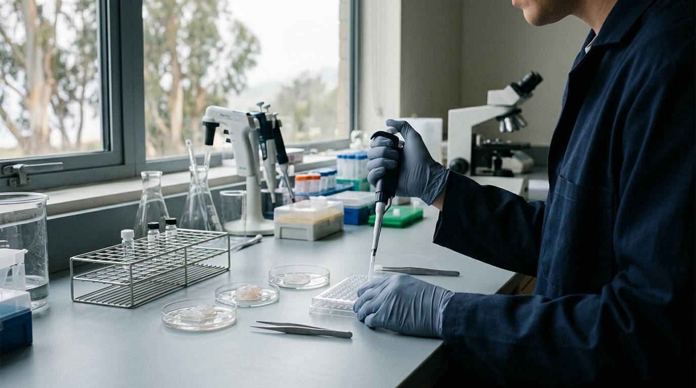
A reduction in wound margins does not prove that damaged tissue has successfully healed. In preclinical cutaneous repair studies, visual wound closure frequently masks incomplete structural recovery or a defective epidermal barrier. Treating wound area as a universal metric can lead investigators to draw flawed conclusions about tissue regeneration.
Careful evaluation requires matching specific scientific questions to appropriate animal systems, injury types, and quantitative readouts. This guide analyzes how preclinical models represent acute trauma, burns, and chronic microbial infections. It details the physiological mechanisms governing closure across species and highlights the diagnostic tools required to verify true tissue restoration. Investigators exploring literature in recovery and regenerative research must understand these parameters to properly evaluate published data.
Differentiate Rodent Contraction From Human Re-Epithelialization
A common challenge in preclinical cutaneous studies involves anatomical variation between species. Rodents possess the panniculus carnosus, a subcutaneous striated muscle layer beneath the loose skin. Humans lack this anatomical layer. Because of this muscle layer, unsplinted full-thickness wounds in rodents close primarily through rapid wound contraction.
Human wound closure relies primarily on re-epithelialization and granulation tissue formation rather than rapid contraction. When an unsplinted rodent wound contracts, the margins pull together rapidly. This mechanism bypasses the slower processes of keratinocyte migration and cellular proliferation that characterize human repair. Evaluating a candidate compound in an unsplinted rodent model can therefore misrepresent how the agent influences human tissue repair.
To address this challenge, researchers developed the splinted full-thickness excisional model in mice. In this protocol, investigators create dorsal excisional wounds and secure rigid silicone rings around the periphery. These splints mechanically resist the inward pull of the panniculus carnosus. As a result, the splinted model minimizes contraction and forces the injury to heal predominantly through re-epithelialization.
When reviewing study designs, checking whether rodent excisional models utilize splinting is essential. Without mechanical splinting, reductions in wound size reflect rodent-specific musculature dynamics rather than true epithelial resurfacing. Researchers reviewing papers should consult structured guides on how to read a research paper to verify whether splints were used in mouse wound studies.
Evaluate Impaired Repair In Diabetic And Microbial Challenge Systems
Standard acute wounds in healthy animals heal quickly, which makes them poor representations of human chronic wounds. Chronic human ulcers typically involve underlying metabolic dysfunction, continuous inflammation, and persistent bacterial colonization. To reproduce these hostile environments, investigators often use genetically altered animals combined with microbial inoculation.
A primary example involves the diabetic mouse wound model using genetically diabetic db/db mice. In one published study, researchers placed six-millimeter dorsal punch biopsy wounds on db/db mice. Two days after wounding, the researchers inoculated the injuries with Pseudomonas aeruginosa biofilms and covered them with semi-occlusive dressings for two weeks. The study followed the animals across a 28-day observation period.
The experimental challenge produced clear differences in wound resolution. Most uninfected control wounds in the diabetic mice achieved re-epithelialization by day 28. In contrast, none of the wounds challenged with P. aeruginosa biofilms achieved closure within that timeframe. Histological evaluation revealed extensive inflammatory cell infiltration, tissue necrosis, and epidermal hyperplasia adjacent to the wound margins.
These histological findings confirmed that bacterial biofilms prevent normal repair and sustain a destructive inflammatory state. When evaluating chronic wound research, verifying that investigators tracked tissue morphology alongside visual measurements is essential. Relying solely on gross macroscopic inspection can miss deep necrosis and cellular pathology beneath the surface. Adhering to rigorous research standards requires analyzing both microscopic histology and physical wound margins.
Separate Surface Planimetry From Functional Barrier Recovery
Planimetry measures the surface area of an open wound across progressive time points. While planimetry provides a simple quantitative measurement, it cannot confirm whether the newly formed epidermis functions correctly. A wound can appear closed by two-dimensional photographic analysis while remaining functionally compromised.
This divergence was documented in a porcine full-thickness burn model. Researchers created eight full-thickness burns symmetrically on the dorsum of female domestic white pigs. Three days post-burn, the investigators inoculated the burn sites with Pseudomonas aeruginosa. They tracked repair over time using laser speckle imaging, high-resolution ultrasound, and transepidermal water loss measurements alongside wound area planimetry.
The porcine burn study revealed that macroscopic wound margins could shrink while transepidermal water loss remained significantly elevated. Elevated water loss demonstrates that the stratum corneum has not restored its hydrophobic seal. Despite the visual appearance of closure, the underlying tissue remains permeable and vulnerable to environmental insults.
Porcine skin shares key physiological characteristics with human skin, including dermal thickness, hair follicle distribution, and repair kinetics. Because pigs heal predominantly through re-epithelialization rather than contraction, they serve as a valuable model for human cutaneous thermal trauma. However, the study confirms that planimetric wound size and functional barrier restoration are independent parameters that cannot substitute for one another.
Account For Laboratory Standards And Study Boundaries
Translating preclinical wound outcomes to broader biological conclusions requires strict methodological boundaries. Animal models provide controlled settings to study specific biochemical pathways, but each model possesses distinct biological limitations. Rodent splints reduce contraction, yet mouse skin still differs from human skin in terms of epidermal thickness and immunological responses.
Similarly, porcine burn models offer human-relevant architecture, but they are technically demanding and ethically constrained by sample size limitations. Experimental biofilm models use defined, lab-grown bacterial strains that do not fully match the polymicrobial communities found in clinical wounds. A model shows how an experimental intervention behaves under isolated, controlled parameters, not how it functions across heterogeneous biological settings.
Laboratory quality controls also require distinct interpretation from in vivo performance. In experimental settings, analytical testing such as chromatography and mass spectrometry confirms the identity, purity, and composition of research peptides. A Certificate of Analysis confirms only that a specific lot matches its chemical specifications under verified analytical methods.
A laboratory test cannot establish biological efficacy, tissue absorption, or safety in a living organism. Researchers must never treat analytical purity as evidence of biological performance in wound repair. Scientific integrity requires maintaining a strict separation between chemical verification in the laboratory and biological activity observed across experimental wound models.
Key Takeaways
- Unsplinted rodent wounds close primarily through contraction mediated by the panniculus carnosus, a muscle layer that humans do not possess.
- Securing silicone splints around full-thickness mouse wounds mechanically limits contraction and forces repair to proceed through re-epithelialization.
- In diabetic mouse models, inoculation with Pseudomonas aeruginosa biofilms arrests wound closure and produces severe tissue necrosis and inflammatory infiltration.
- Macroscopic wound closure does not guarantee that the skin barrier is restored, as shown by persistent water loss in porcine burn injuries.
- Chemical purity reports verify only the physical contents of a specific laboratory sample and do not establish biological safety or healing activity.
Careful wound-healing research requires selecting experimental models that reflect target human repair mechanisms while measuring cellular architecture and barrier integrity alongside visual surface closure.
Sources
Introducing / CaliLean Field Notes
A little more
perspective.
An editorial letter for curious minds. Thoughtful reading, questions worth keeping and the sources behind them.
Explore Field Notes Join the list for the first edition

