
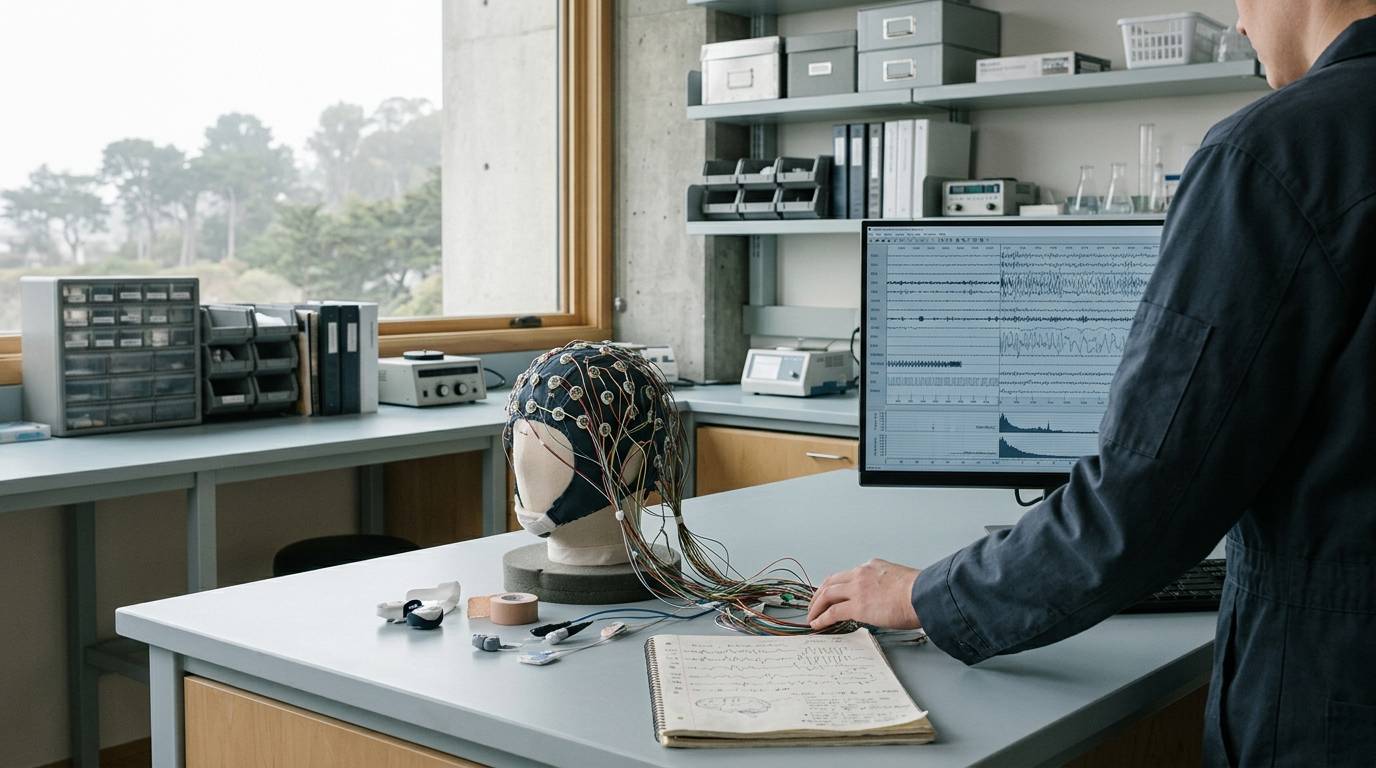
Sleep research often encounters a fundamental challenge of definition. Stating that an intervention improved sleep does not describe a single biological outcome. Sleep is not a uniform physiological block, nor is it an isolated metric. Instead, sleep represents an orchestrated series of physiological states across distinct neurological domains. When researchers assess an intervention, measuring sleep requires specifying precisely which component is under observation.
This resource examines the primary operational domains of sleep research. It clarifies how quantity, continuity, initiation, architecture, and subjective perception diverge across preclinical and clinical paradigms. It also examines why endpoint selection fundamentally alters conclusions in neuropeptide studies and brain, sleep, and cognitive peptide research.
What Distinguishes Sleep Quantity, Continuity, and Perceived Quality?
Sleep cannot be reduced to a single score. In neuroscience research, investigators divide sleep into at least five primary operational domains. These domains include quantity, continuity, initiation, architecture, and subjective perception. While these domains are physiologically related, they frequently move in opposite directions.
Quantity represents the total duration of sleep generated over a defined window. The most common index is total sleep time, which aggregates all scored epochs of sleep. Quantity answers how much sleep occurred. However, it reveals nothing about whether that sleep was fragmented or consolidated.
Continuity describes the stability and maintenance of the sleep episode across time. Key parameters include wake after sleep onset, the number of nocturnal awakenings, and sleep efficiency. A subject may obtain a large quantity of sleep that remains broken by frequent brief arousals. Conversely, a subject may experience brief sleep that is tightly consolidated.
Sleep initiation evaluates the transition from wakefulness to sleep. Sleep onset latency serves as the primary metric for this domain. An intervention may accelerate sleep initiation without altering subsequent continuity or overall duration. Conflating faster initiation with longer sleep misrepresents the biological target of the compound.
Sleep architecture describes the structural distribution of physiological sleep states across the recording window. In polysomnography protocols, this reflects the time spent in non-rapid eye movement and rapid eye movement sleep. Architecture captures whether sleep exhibits standard physiological organization and expected cyclic transitions.
Perceived quality addresses subjective experience and reported daytime restoration. Research literature highlights that sleep quality is an inherently multidimensional construct. It cannot be reduced to objective sleep duration. Subjective quality reflects perceived depth, continuous rest, and morning alertness, which often decouple from physiological recordings.
When a research team evaluates a compound, attributing an effect to sleep quality requires assessing subjective experience directly. If an investigator only records an increase in sleep duration, reporting improved quality overstates the findings. Researchers must report findings within the precise domain evaluated by their experimental tools.
How Are Core Objective Metrics Defined and Calculated in Laboratory Protocols?
Objective sleep metrics require standardized operational definitions to allow cross-study comparison. Slight differences in scoring rules or mathematical denominators can substantially alter the apparent outcome of a trial. Laboratory investigations typically derive these variables from continuous electrophysiological recordings.
Total sleep time represents the aggregate duration of all scored sleep states during an active recording period. Technical guidance defines total sleep time as the sum of non-rapid eye movement stages and rapid eye movement sleep across scored epochs. Epochs are traditionally scored in sequential 30-second windows. Total sleep time does not indicate whether sleep was restorative or properly aligned with circadian rhythms.
Sleep onset latency measures the interval required for a subject to transition from wakefulness to sleep. The starting point is typically defined as lights out, bedtime, or the formal beginning of a sleep opportunity. The endpoint is defined by a standardized scoring rule, such as the appearance of the first epoch of any sleep state. Because some laboratories define onset as the first three consecutive epochs of stage N1 or the first epoch of stage N2, researchers must disclose their exact rule.
Wake after sleep onset quantifies the total duration of wakefulness occurring between sleep onset and final awakening. This variable serves as a direct indicator of sleep maintenance and structural continuity. An operational definition must define both the initial onset rule and the final awakening threshold. A reduction in wake after sleep onset demonstrates improved consolidation, even when total sleep time remains unchanged.
Sleep efficiency is calculated by dividing total sleep time by time in bed, then multiplying by one hundred. This metric depends equally on the sleep numerator and the time-in-bed denominator. If an experimental protocol restricts time in bed, sleep efficiency will appear higher even if total sleep time declines. Research reports should always specify whether the denominator represents total time in bed, the prescribed sleep opportunity, or another interval.
Awakening count and arousal index both relate to continuity, but they quantify distinct physiological phenomena. An awakening typically involves an epoch-level transition from sleep to behavioral wakefulness lasting at least thirty seconds. A microarousal represents an abrupt shift in electroencephalographic frequency lasting as little as three seconds, often without conscious awakening. A drug that decreases macro-awakenings might leave the microarousal index elevated, leaving sleep fragmenting processes active.
Why Does Sleep Architecture Matter Beyond Total Sleep Time?
Assessing total sleep time alone provides an incomplete picture of physiological sleep. Polysomnography protocols categorize sleep into discrete states based on electroencephalogram, electrooculogram, and electromyogram signals. The American Academy of Sleep Medicine established standardized staging criteria that divide sleep into non-rapid eye movement stages and rapid eye movement sleep.
Stage N1 represents the initial transition from alpha-dominant wakefulness to light sleep. It is characterized by low-amplitude, mixed-frequency electroencephalographic activity and slow rolling eye movements. Stage N2 represents deeper baseline sleep characterized by the appearance of sleep spindles and K-complexes. Spindles reflect thalamocortical oscillations that shield the cortex from external sensory inputs.
Stage N3 represents slow-wave sleep, which combines the former stages S3 and S4 from older classification manuals. Stage N3 is defined by high-amplitude delta waves occurring at low frequencies across frontal cortical leads. This stage correlates with metabolic clearance, growth hormone secretion, and synaptic downscaling. Rapid eye movement sleep features low-voltage mixed-frequency brain activity, muscle atonia, and rapid saccadic eye movements.
Changes in architecture reveal pharmacological mechanisms that overall duration metrics obscure. For instance, classic sedative-hypnotic compounds often increase total sleep time while simultaneously suppressing stage N3 slow-wave sleep and rapid eye movement sleep. Such compounds extend duration primarily by expanding stage N2 sleep. Evaluating only total sleep time would categorize these compounds as broadly sleep-promoting, masking their disruptive effects on restorative slow-wave architecture.
Quantitative spectral analysis provides an even deeper assessment of sleep architecture beyond categorical staging. Researchers evaluate slow-wave activity by measuring electroencephalographic power density within the delta frequency band during non-rapid eye movement sleep. Slow-wave activity reflects homeostatic sleep pressure that accumulates during prior wakefulness and dissipates across the sleep period. Measuring changes in delta power reveals whether an intervention alters the biological intensity of sleep, which categorical epoch staging cannot capture.
A change in sleep stage distribution should not be automatically categorized as beneficial. While theoretical models associate stage N3 with somatic restoration and rapid eye movement with memory consolidation, changing their proportions can produce unintended cognitive consequences. A descriptive reporting framework remains the most accurate standard. Researchers should describe the observed shifts in minutes or stage percentages, avoiding unsupported conclusions about sleep depth.
How Do Measurement Methods Shape Sleep Data and Introduce Potential Confounds?
The selection of measurement technology fundamentally shapes the resulting sleep dataset. Neuroscience investigations deploy three primary measurement modalities: polysomnography, actigraphy, and subjective self-report instruments. Each approach operates with specific mechanical assumptions, analytical strengths, and systematic failure points.
Polysomnography represents the gold-standard objective modality for assessing sleep physiology. By synchronizing electroencephalography, electromyography, and electrooculography, polysomnography directly tracks neurophysiological state transitions. However, the modality carries significant logistical burdens. It requires specialized sensor montages, laboratory recording spaces, and laborious scoring by trained technical personnel.
The artificial nature of the laboratory environment introduces experimental confounds. The classic first-night effect describes altered sleep architecture and heightened fragmentation caused by unfamiliar laboratory settings and sensor arrays. Researchers frequently discard data from the initial recording night to allow subject habituation. Portable polysomnography systems mitigate this confound by recording within home environments, though they carry higher rates of electrode displacement and signal loss.
Actigraphy provides a continuous, non-invasive alternative by capturing movement via wrist-worn accelerometers. Algorithms process raw acceleration signals into activity counts, inferring sleep during intervals of prolonged immobility. Actigraphy allows researchers to gather multi-week recordings within natural living conditions at relatively low cost. However, actigraphy measures gross physical movement rather than central sleep physiology.
Because actigraphy infers sleep purely from lack of movement, it exhibits predictable systematic errors. In populations characterized by high sleep fragmentation or severe insomnia, actigraphy frequently misclassifies quiet wakefulness as sleep. When sleep efficiency falls below 73%, the concordance between actigraphy and polysomnography deteriorates substantially. Passive movement in bed or motor conditions like periodic limb movements will conversely cause algorithms to misclassify true sleep as wakefulness.
Subjective measurement tools include daily sleep diaries and standardized questionnaires like the Pittsburgh Sleep Quality Index. These instruments capture the subject's internal experience of sleep, perceived latency, and daytime functional impairment. While these variables directly measure patient-centered outcomes, they correlate poorly with objective polysomnographic recordings.
This mismatch between subjective reports and objective recordings is known as sleep state misperception. Individuals with insomnia often report zero sleep despite electroencephalographic records demonstrating several hours of continuous sleep. Conversely, healthy individuals frequently overestimate their sleep duration by failing to recall brief nocturnal arousals. Discrepancies between actigraphy, polysomnography, and diaries do not necessarily indicate measurement failure, but rather demonstrate that each tool captures a distinct biological or experiential axis.
What Can Preclinical Peptide Models Tell Us About Sleep Regulation?
Preclinical peptide research offers valuable insight into the central circuits governing arousal and somnolence. Neuropeptides synthesized within distinct hypothalamic nuclei coordinate the transitions between wakefulness, non-rapid eye movement sleep, and rapid eye movement sleep. Understanding these circuits helps researchers apply appropriate rigorous research methodology frameworks to experimental designs.
The orexin system, also known as the hypocretin system, provides a well-documented model of neuropeptide sleep-wake regulation. Orexin-A and orexin-B are synthesized by a discrete cluster of neurons located in the lateral hypothalamic area. These neurons project extensively to monoaminergic and cholinergic arousal centers throughout the brainstem and basal forebrain. These targets include the locus coeruleus, the tuberomammillary nucleus, and the dorsal raphe nuclei.
Activation of orexin receptors stabilizes wakefulness and prevents inappropriate transitions into sleep. Preclinical rodent studies demonstrate that central administration of orexin-A produces robust behavioral arousal. Intracerebroventricular injection of orexin-A in rodents suppresses both non-rapid eye movement sleep and rapid eye movement sleep while driving significant increases in electroencephalographic wakefulness. In contrast, blocking this signaling pathway yields opposite physiological outcomes.
Dual orexin receptor antagonists and selective orexin receptor-2 antagonists promote sleep by inhibiting this endogenous wake drive. In rodent models, orexin receptor antagonists consistently increase both non-rapid eye movement and rapid eye movement sleep duration. These pharmacodynamic effects highlight why peptide signaling must be evaluated with endpoint precision. Administering an endogenous peptide ligand and administering a receptor antagonist alter the sleep-wake network in fundamentally different directions.
Preclinical sleep protocols require meticulous control over experimental variables. In one standardized rodent design, mice received intracerebroventricular microinjections of orexin-A at doses of 0.3, 1, or 3 nanomoles or a vehicle control. Injections were administered exactly three hours into the light phase using a randomized crossover design with a three-to-four-day washout interval. Continuous electroencephalographic and electromyographic data were acquired across an eight-hour post-injection observation window.
Failing to report such procedural details compromises scientific reproducibility. Rodents are nocturnal animals that exhibit polyphasic sleep patterns, concentrating their rest during the light phase. An intervention administered during the light phase tests a compound's ability to disrupt consolidated sleep or promote wakefulness. Administering the same compound during the dark phase tests its capacity to overcome active wake drives and induce somnolence.
Peptide sleep outcomes also depend heavily on underlying pathological models. In APP/PS1 transgenic mouse models of Alzheimer's disease, researchers observed marked alterations in orexinergic tone and sleep architecture. Wakefulness increased by 42.9% during the twelve-hour light phase and by 12.1% across the entire twenty-four-hour recording period. Simultaneously, non-rapid eye movement sleep decreased by 16.9% during the dark phase and 10.3% over twenty-four hours.
These observations reflect model-specific neuropathology and cannot be directly translated into human clinical predictions. Rodent sleep architecture differs substantially from human monophasic sleep. Mice transition between brief bouts of wake, non-rapid eye movement, and rapid eye movement sleep every few minutes. Translating a rodent electroencephalographic signal into a clinical outcome carries substantial translational risk, as detailed in discussions of evaluating translational risk in preclinical studies.
Why Do Melatonin and Chronobiotic Studies Highlight Methodological Divergence?
Melatonin research provides a clear example of how endpoint and measurement selection dictate study conclusions. While melatonin is a small-molecule neurohormone rather than a peptide, its literature illustrates methodological divergence across sleep outcome domains. Researchers must examine these patterns to avoid confusing chronobiotic shifts with direct sedative mechanisms.
Melatonin acts primarily through MT1 and MT2 G-protein coupled receptors located in the suprachiasmatic nucleus of the anterior hypothalamus. Its endogenous secretion conveys circadian phase information, signaling biological night rather than directly inducing central nervous system sedation. Consequently, testing melatonin against traditional hypnotic endpoints often yields mixed or contradictory findings depending on the tools used.
A comprehensive meta-analysis evaluating melatonin across primary sleep disorders illustrated this divergence. When researchers pooled subjective questionnaire data, melatonin demonstrated a statistically significant increase in total sleep time. However, when the analysis isolated objective polysomnographic recordings, the increase in total sleep time was no longer statistically significant. Melatonin reliably reduced sleep onset latency across both modalities, but its effect on duration differed between assessment methods.
This pattern demonstrates that subjective ratings and objective polysomnographic recordings capture different dimensions of the sleep episode. A subject may experience an easier transition into sleep and report feeling more rested the following morning. Yet their total electrophysiological sleep time may remain identical to placebo conditions. If an author describes this outcome as an objective increase in sleep duration, the statement misrepresents the data.
Chronobiotic studies also show that administration timing alters the primary endpoint. Administering a chronobiotic compound during the biological day shifts the circadian phase of the central pacemaker, altering subsequent sleep timing. Administering the exact same dose during the biological night produces negligible phase-shifting effects and minimal hypnotic action. Investigators must separate phase-shifting outcomes from direct somnogenic properties when evaluating sleep-active compounds.
What Methodological Pitfalls Distort Findings in Sleep Outcome Research?
Sleep outcome literature is frequently compromised by avoidable methodological errors and imprecise interpretations. Identifying these pitfalls enables researchers to construct stronger protocols and evaluate published literature with appropriate skepticism. The most common distortions involve overgeneralization, baseline variability, operational ambiguity, and analytical multiplicity.
Conflating Individual Metric Shifts With Generalized Sleep Improvement
The most pervasive error in sleep research is labeling a shift in a single parameter as generalized sleep improvement. A reduction in sleep onset latency demonstrates only that sleep initiation occurred more rapidly. It does not establish that the sleep was continuous, structurally sound, or perceived as restorative. Similarly, an isolated increase in total sleep time does not indicate that the subject received healthy sleep.
If an intervention increases total sleep time but induces sleep fragmentation, calling the overall outcome an improvement is inaccurate. Researchers must identify the precise metric that changed, report its mathematical direction, and avoid broad evaluative claims. Subjective restoration should only be reported if validated patient-reported outcome measures were directly administered.
Overlooking Multi-Night Variability and Laboratory Habituation
Sleep parameters exhibit substantial variability from night to night within the same individual. In a home-monitoring study evaluating 94 participants across 470 consecutive recording nights, night-to-night variability was high for several core metrics. Wake after sleep onset, sleep onset latency, persistent sleep latency, and time spent in stages N2 and N3 varied widely across nights. In contrast, total sleep time and overall sleep efficiency showed higher intra-individual stability.
Relying on a single night of polysomnography to characterize an individual's sleep profile introduces severe sampling error. A single recording cannot capture typical baseline variability or distinguish night-to-night fluctuations from true pharmacological effects. For subjective diary assessments, research indicates that at least six consecutive recording nights are required to establish reliable baseline estimates. Multi-night monitoring designs are essential for generating dependable sleep data.
Inconsistent Sleep Onset Definitions and Unspecified Denominators
Sleep onset latency appears straightforward, yet its calculation varies widely across laboratory protocols. One protocol may define sleep onset as the first thirty-second epoch of any sleep stage, including stage N1. Another protocol may require three consecutive epochs of stage N1 or the appearance of a single epoch of stage N2. These disparate criteria yield markedly different latency values from the exact same electroencephalographic trace.
Sleep efficiency calculations suffer from similar ambiguity. Some studies divide total sleep time by total time spent in bed, while others divide total sleep time by the designated lights-out interval. If an experimental protocol keeps a subject in bed for an extra hour after awakening, sleep efficiency drops automatically even if sleep duration was maintained. Authors must report both the numerator and the denominator used to calculate efficiency.
Equating Sleep Duration With Sleep Consolidation
An intervention can expand total sleep time while degrading sleep continuity. In a rodent pharmacological study testing dual orexin receptor antagonists, acute drug administration increased total rapid eye movement and non-rapid eye movement sleep during the dark phase. However, electrophysiological analysis revealed that sleep fragmentation remained elevated, and non-rapid eye movement delta power was unaffected.
If the investigators had only tracked total sleep duration, they would have concluded that sleep improved uniformly. By tracking continuity metrics alongside spectral power, they showed that the expanded sleep remained fragmented and lacked increased slow-wave intensity. Quantity improvements should always be paired with continuity metrics and spectral power analyses to provide a complete picture of sleep stability.
Multiplicity and Selective Endpoint Reporting
Modern polysomnography generates dozens of distinct outcome variables per recording session. These include total sleep time, latency to each stage, awakenings, microarousal indices, stage percentages, and spectral power bands. When an investigator measures dozens of variables across multiple time windows, false-positive findings emerge through chance alone.
Selective reporting occurs when authors highlight the few variables that achieved statistical significance while omitting the dozens that showed no change. To ensure methodological rigor, protocols must pre-specify primary and secondary endpoints prior to trial execution. When testing multiple secondary endpoints, investigators must apply formal correction procedures, such as false-discovery-rate control or Bonferroni adjustments.
How Should Researchers Select and Report Sleep Endpoints to Ensure Reproducibility?
Standardized reporting standards are essential for improving reproducibility in sleep outcome research. When describing sleep experiments, researchers should follow a structured reporting framework that defines the exact scope and limitations of their data.
Investigators must first identify the precise operational domain under evaluation, explicitly categorizing the endpoint as quantity, continuity, initiation, architecture, timing, or perceived quality. Next, the exact metric and its measurement units must be disclosed, such as minutes for wake after sleep onset or percentages for sleep efficiency. Authors should define the operational boundaries of the metric, including lights-out parameters, sleep-onset scoring thresholds, and the denominator used for efficiency.
The technical measurement modality must be clearly stated. When reporting polysomnography, authors must document electrode montages, epoch lengths, and the specific scoring manual applied. When reporting actigraphy, the physical device model, sampling frequency, and scoring algorithm must be detailed. When subjective outcomes are gathered, the validated questionnaire or diary format should be named.
Study designs should report the total duration of the recording period and the number of consecutive nights evaluated. Researchers must describe the experimental population in detail, including species, strain, age, sex, and underlying disease models. The circadian timing of drug delivery relative to the ambient light-dark cycle must be documented, along with the precise post-dose observation window analyzed.
Finally, researchers must distinguish between receptor binding and downstream biological function. Demonstrating that a peptide binds to an orexin, melatonin, or neuropeptide receptor does not prove that it will alter sleep outcomes in an intact animal. The distinction between receptor binding and downstream biological effects remains a critical dividing line in preclinical pharmacology.
Investigators must also maintain rigorous standards when characterizing peptide materials used in sleep studies. A Certificate of Analysis or independent laboratory test establishes the chemical identity and purity of a specific submitted sample. It confirms whether the analyzed aliquot contains the intended peptide sequence and quantifies residual counterions or synthesis impurities. Analytical validation frameworks are detailed in resources covering laboratory analytical standards.
However, an analytical testing record does not predict biological activity, in vivo receptor affinity, or physiological sleep outcomes. A Certificate of Analysis confirms chemical purity, but it cannot confirm how that compound will behave in an electrophysiological recording chamber. Researchers must maintain this distinction to prevent chemical verification from being confused with biological efficacy.
Frequently Asked Questions About Sleep Metrics and Peptide Models
Why do actigraphy and polysomnography frequently yield discordant wake after sleep onset values?
Actigraphy infers sleep and wakefulness exclusively from physical acceleration. If an experimental subject lies quietly awake in bed without moving, the actigraphy algorithm will register the absence of movement as sleep. Consequently, actigraphy frequently underestimates wake after sleep onset, particularly in individuals with severe sleep fragmentation or insomnia. Polysomnography directly tracks central electroencephalographic activity, identifying cortical arousal even when the subject remains physically motionless.
How does circadian phase timing alter the behavioral response to neuropeptide administration?
The central nervous system exhibits pronounced circadian fluctuations in baseline receptor expression, endogenous ligand tone, and arousal circuit excitability. For example, administering an orexin-A receptor agonist during the light phase tests its capacity to disrupt established sleep in nocturnal rodents. Administering the identical compound during the active dark phase produces less noticeable behavioral changes because endogenous arousal circuits are already operating at peak capacity. Researchers must align the timing of compound administration with the specific circadian hypothesis being tested.
Can sleep architecture improvements be inferred solely from an increase in total sleep time?
An increase in total sleep time provides no structural information regarding sleep architecture. An intervention can substantially increase total sleep time by extending light stage N2 sleep while simultaneously suppressing stage N3 slow-wave sleep and rapid eye movement sleep. Architecture can only be evaluated by directly scoring individual sleep stages via continuous electrophysiological recordings. Asserting that sleep architecture improved requires demonstrating appropriate stage distributions and normal cyclic transitions across the sleep period.
What distinguishes microarousals from overt awakenings in electroencephalographic sleep recordings?
An awakening involves a clear behavioral transition from sleep to wakefulness that typically persists for at least thirty seconds, scoring as an epoch of wake. A microarousal represents an abrupt, temporary shift in electroencephalographic frequency lasting between three and fifteen seconds. Microarousals reflect transient autonomic and cortical activation without conscious behavioral awakening. A compound may successfully suppress overt awakenings while leaving microarousal frequency elevated, maintaining fragmented sleep that compromises daytime recovery.
Sources
- Meta-analysis: melatonin for the treatment of primary sleep disorders
- Sleep Classification According to AASM and Rechtschaffen & Kales
- Orexin A peptidergic system: comparative sleep behavior, morphology and population in brains between wild type and Alzheimer’s disease mice
- Assessment of Subjective and Objective Sleep Quality ...
- Effects of lemborexant and zolpidem on sleep ...
- Objective multi-night sleep monitoring at home: variability ...
- Effect of daridorexant on sleep architecture in patients with chronic insomnia disorder: a pooled post hoc analysis of two randomized phase 3 clinical studies
Introducing / CaliLean Field Notes
A little more
perspective.
An editorial letter for curious minds. Thoughtful reading, questions worth keeping and the sources behind them.
Explore Field Notes Join the list for the first edition

