
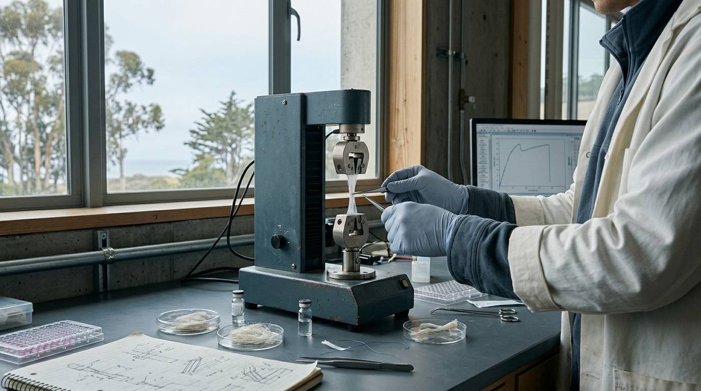
Many researchers struggle to determine whether higher collagen content in healing connective tissue indicates genuine structural repair. A routine laboratory query centers on why a tendon with dense collagen staining often fails under modest physical force. This guide provides an evidence-based answer by examining how mechanical loading affects matrix organization, cellular alignment, and experimental endpoints in tendon repair models.
Scope of Connective Tissue Repair Analysis
Investigating tendon and ligament healing requires distinguishing between crude protein accumulation and functional tissue regeneration. Collagen production alone does not verify that a healing tendon has regained structural competence.
Dense connective tissues rely on parallel collagen fiber bundles to resist tensile loads. When an injury disrupts this matrix, the reparative cascade produces a provisional scaffold that often lacks native hierarchy. Measuring total extracellular matrix content without assessing alignment or architecture obscures whether the tissue has repaired properly or formed a disorganized scar. Researchers can review structural assessment criteria in our research methods library to avoid common measurement pitfalls.
Evidence Context in Preclinical Loading Models
Evaluating tendon repair biology depends heavily on controlled animal models, where mechanical forces can be modified systematically during the healing process. Controlled human trials rarely allow invasive matrix sampling or precise immobilization protocols during early post-injury windows.
Preclinical animal studies offer the resolution needed to compare histological outcomes under distinct physical environments. However, animal data cannot be assumed to translate directly into human clinical timelines. Understanding where these models fit on the translational spectrum is essential for interpreting matrix outcomes, as discussed in our peptide research evidence levels guide. The scientific evidence reviewed here derives strictly from controlled preclinical rodent research rather than human observational or clinical trials.
Biological Study Models and Experimental Conditions
Experimental investigations of mechanical loading frequently rely on surgical transection models. In a published study on rat Achilles tendon healing, female rats underwent complete tendon transection to assess matrix remodeling across varied mechanical environments, according to research published in Scientific Reports.
The investigators established three distinct experimental conditions to isolate the role of tension during healing:
- Full loading, where animals maintained normal cage activity and weight-bearing.
- Reduced loading, where physical tension was partially decreased.
- Minimal loading, where mechanical forces on the transected limb were substantially eliminated.
The experimental period examined the initial three weeks of repair. This interval captures the critical shift from acute inflammation to early matrix synthesis and structural reorganization.
Extracellular Matrix Remodeling and Cellular Alignment
The healing response in dense fibrous connective tissue depends on precise spatial organization. Fibroblasts and tenocytes align along vectors of tension, secreting collagen fibrils that gradually pack into functional fascicles.
When mechanical loading is present, physical tension acts as a directional cue for cellular behavior. Tissue sections stained with hematoxylin and eosin, along with immunohistochemical markers targeting collagen type I, collagen type III, elastin, and CD31, allow researchers to evaluate matrix quality. Without appropriate tension, cells produce matrix components without establishing the parallel orientation required for directional tensile strength. For related background on tissue regeneration, explore our recovery, repair and regenerative research section.
Research Findings Under Variable Loading States
The rat Achilles transection study revealed distinct differences between tissue composition and structural organization across loading conditions. Fully loaded tendons demonstrated significantly greater cellular alignment and matrix organization during early healing. By three weeks post-injury, hematoxylin and eosin staining confirmed that increased mechanical loading produced an organized, directional extracellular matrix.
In contrast, minimally loaded tendons presented an unexpected histochemical profile. These shielded tissues exhibited higher unadjusted staining intensity for collagen type I, collagen type III, and elastin compared to fully loaded controls.
Despite having higher staining intensity for these matrix proteins, the minimally loaded tendons were physically smaller and thinner. The newly synthesized matrix also displayed an unorganized architectural pattern. Researchers tracking only staining intensity would have mischaracterized the unloaded tissue as superior, overlooking its reduced size and lack of linear structure. This divergence emphasizes the conceptual gap between molecular presence and functional architecture, a topic examined further in our analysis of experimental models and biological effects.
Methodological Limitations and Experimental Unknowns
Histological and immunohistochemical measurements have inherent technical limitations in biomechanical research. In the rat Achilles transection experiment, the authors noted an important experimental boundary: the study did not perform mechanical testing.
Because tensile strength, stiffness, and ultimate failure load were not directly measured, the staining results cannot establish which group possessed stronger tendons. Higher staining intensity cannot be equated with load-bearing capacity, nor does cellular alignment prove functional stiffness without physical verification.
Additionally, the investigation examined only female rats within a three-week observation window. These results cannot be generalized to male subjects, longer healing periods, or other anatomical structures without dedicated experimental verification. A careful review of these analytical limits is standard practice when evaluating preclinical data, as detailed in our guide on how to read a research paper.
Analytical Context for Research Materials
Laboratory research on tendon biology depends on verified chemical reagents, peptides, and culture media. When reviewing an analytical report or Certificate of Analysis for research materials, investigators must understand what the testing establishes.
An analytical report confirms the chemical identity, purity, and molecular mass of the specific sample tested. It does not establish biological activity, tissue integration, or mechanical performance in living animal models. Analytical data verifies that an experimental compound matches its expected chemical profile before use in an assay. It does not replace empirical testing in cell lines or animal tissues.
Protocol Considerations for Future Research
Designing rigorous tendon repair studies requires selecting endpoints that assess structural architecture alongside protein accumulation. Researchers planning upcoming experimental protocols can apply the following evaluation checklist:
- Pair staining intensity measurements with cross-sectional area calculations to prevent misinterpreting protein concentration in smaller, atrophic tissues.
- Measure cellular and collagen alignment vectors rather than relying entirely on bulk protein quantification.
- Include mechanical testing endpoints, such as ultimate tensile strength and elastic modulus, alongside histological staining whenever tissue size allows.
- Document the exact loading state of the animal model to avoid confounding structural variables during matrix evaluation.
Sources
Introducing / CaliLean Field Notes
A little more
perspective.
An editorial letter for curious minds. Thoughtful reading, questions worth keeping and the sources behind them.
Explore Field Notes Join the list for the first edition

